Book - The Elements of Embryology - Chicken 2
| Embryology - 28 Apr 2024 |
|---|
| Google Translate - select your language from the list shown below (this will open a new external page) |
|
العربية | català | 中文 | 中國傳統的 | français | Deutsche | עִברִית | हिंदी | bahasa Indonesia | italiano | 日本語 | 한국어 | မြန်မာ | Pilipino | Polskie | português | ਪੰਜਾਬੀ ਦੇ | Română | русский | Español | Swahili | Svensk | ไทย | Türkçe | اردو | ייִדיש | Tiếng Việt These external translations are automated and may not be accurate. (More? About Translations) |
Foster M. Balfour FM. Sedgwick A. and Heape W. The Elements of Embryology (1883) Vol. 1. (2nd ed.). London: Macmillan and Co.
| Historic Disclaimer - information about historic embryology pages |
|---|
| Pages where the terms "Historic" (textbooks, papers, people, recommendations) appear on this site, and sections within pages where this disclaimer appears, indicate that the content and scientific understanding are specific to the time of publication. This means that while some scientific descriptions are still accurate, the terminology and interpretation of the developmental mechanisms reflect the understanding at the time of original publication and those of the preceding periods, these terms, interpretations and recommendations may not reflect our current scientific understanding. (More? Embryology History | Historic Embryology Papers) |
A Brief Summary of the Whole History of Incubation
STEP by step the simple two-layered blastoderm described in the previous chapter is converted into the complex organism of the chick. The details of the many changes through which this end is reached will perhaps be rendered more intelligible if we prefix to the special history of them a brief summary of the general course of events from the beginning to the end of incubation.
In the first place, it is to be borne in mind that the embryo itself is formed in the area pellucida, and in the area pellucida alone. The area opaca in no part enters directly into the body of the chick; the structures to which it gives rise are to be regarded as appendages, which sooner or later disappear.
Germinal layers
The blastoderm at starting consists of two layers. Very soon a third layer makes its appearance between the other two. These three layers, known as the germinal layers, the establishment of which is a fact of fundamental importance in the history of the embryo, are called respectively the upper, middle and lower layers, or epiblast, mesoblast and hypoblast. Of these the epiblast and hypoblast constitute the primary layers.
Three similar germinal layers are found in the embryos of all vertebrate and most invertebrate forms, and their history is one of the most important parts of comparative embryology.
The epiblast gives rise to the epidermis, the central and peripheral parts of the nervous system, and to the most important parts of the organs of special sense. The hypoblast is essentially the secretory layer, and furnishes the whole epithelial lining of the alimentary tract and its glands, with the exception of part of the mouth and anus which are lined by the epiblast and are spoken of by embryologists as the stomodceum and proctodceum. Finally the mesoblast is a source from which the whole of the vascular system, the muscular and skeletal system, and the connective tissue of all parts of the body, are developed. It gives in fact origin to the connective-tissue basis both of the skin and of the mucous membrane of the alimentary tract, and to all the structures lying between these two with the exceptions already indicated. It is more especially to be noted that it gives rise to the excretory organs and generative glands.
Formation of the embryo
The blastoderm which at first, as we have seen, lies like a watch-glass over the cavity below, its margin resting on the circular germinal wall of white yolk, spreads, as a thin circular sheet, over the yolk, immediately under the vitelline membrane. Increasing uniformly at all points of its circumference, the blastodermic expansion covers more and more of the yolk, and at last, reaching the opposite pole, completely envelopes it. Thus the whole yolk, instead of being enclosed as formerly by the vitelline membrane alone, comes to be also enclosed in a bag formed by the blastoderm.
It is not however until quite a late period that the complete closing in at the opposite pole takes place ; in fact the extension of the blastoderm must be thought of as going on during the first seven days of incubation.
Both the area opaca and the area pellucida share in this enlargement, but the area opaca increases much more rapidly than the area pellucida, and plays the principal part in encompassing the yolk.
The mesoblast, in that part of the area opaca which is nearest to the area pellucida, becomes the seat of peculiar changes, which result in the formation of bloodvessels. Hence this part of the area opaca is called the vascular area.
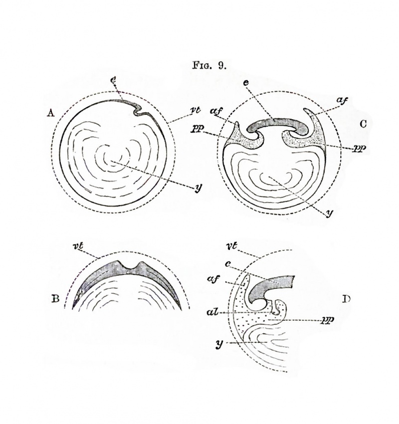
The embryo itself may be said to be formed by a folding off the central portion of the area pellucida from the rest of the blastoderm. At first the area pellucida is quite flat, or, inasmuch as it forms part of the circumference of the yolk, slightly but uniformly curved. Very soon, however, there appears at a certain spot a semilunar groove, at first small, but gradually increasing in depth and extent; this groove, which is represented in section in the diagram (Fig. 9, A\ breaks the uniformity of the level of the area pellucida. It may be spoken of as a tucking in of a small portion of the blastoderm in the form of a crescent. When viewed from above, it presents itself as a curved line (the hinder of the two concentric curved lines in front of A in Fig. 22), which marks the hind margin of the groove, the depression itself being hidden.
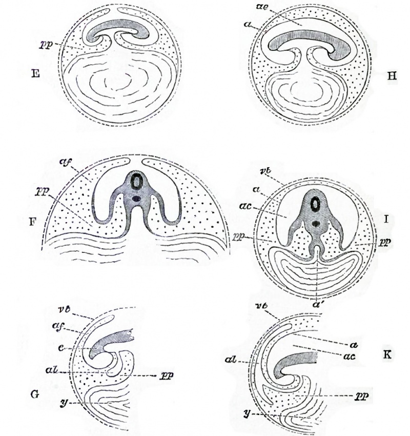
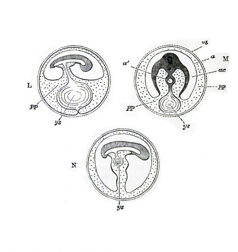
Fig. 9, A to N forms a series of purely diagrammatic representations introduced to facilitate the comprehension of the manner in which the body of the embryo is formed, and of the various relations of the yolk-sac, amnion and allantois.
In all vt is the vitelline membrane, placed, for convenience sake, at some distance from its contents, and represented as persisting in the later stages ; in the actual egg it is in direct contact with the blastoderm (or yolk), and early ceases to have a separate existence. In all e indicates the embryo, pp the general pleuroperitoneal space, of the folds of the amnion proper ; ae or ac the cavity holding the liquor amnii ; al the allantois ; a the alimentary canal ; y or ys the yolk or yolk-sac.
A, which maybe considered as a vertical section taken longitudinally along the axis of the embryo, represents the relations of the parts of the egg at the time of the first appearance of the head-fold, seen on the right-hand side of the blastoderm e. The blastoderm is spreading both behind (to the left hand in the figure), and in front (to right hand) of the head-fold, its limits being indicated by the shading and thickening for a certain distance of the margin of the yolk y. As yet there is no fold on the left side of e corresponding to the head-fold on the right.
B is a vertical transverse section of the same period drawn for convenience sake on a larger scale (it should have been made flatter and less curved). It shews that the blastoderm (vertically shaded) is extending laterally as well as fore and aft, in fact in all directions ; but there are no lateral folds, and therefore no lateral limits to the body of the embryo as distinguished from the blastoderm.
Incidentally it shews the formation of the medullary groove by the rising up of the laminae dorsales. Beneath the section of the groove is seen the rudiment of the notochord. On either side a line indicates the cleavage of the mesoblast just commencing.
In C, which represents a vertical longitudinal section of later date, both head-fold (on the right) and tail-fold (on the left) have advanced considerably. The alimentary canal is therefore closed in, both in front and behind, but is in the middle still widely open to the yolk y below. Though the axial parts of the embryo have become thickened by growth, the body- walls are still thin ; in them however is seen the cleavage of the mesoblast, and the divergence of the somatopleure and splanchnopleure. The splanchnopleure both at the head and at the tail is folded in to a greater extent than the somatopleure, and forms the still wide splanchnic stalk. At the end of the stalk, which is as yet short, it bends outwards again and spreads over the surface of the yolk. The somatopleure, folded in less than the splanchnopleure to form the wider somatic stalk, sooner bends round and runs outwards again. At a little distance from both the head and the tail it is raised up into a fold, a/, a/, that in front of the head being the highest. These are the amniotic folds. Descending from either fold, it speedily joins the splanchnopleure again, and the two, once more united into an uncleft membrane, extend some way downwards over the yolk, the limit or outer margin of the opaque area not being shewn. All the space between the somatopleure and the splanchnopleure, pp } is shaded with dots. Close to the body this space may be called the pleuroperitoneal cavity ; but outside the body it runs up into either amniotic fold, and also extends some little way over the yolk.
D represents the tail end at about the same stage on a more enlarged scale, in order to illustrate the position of the allantois al (which was for the sake of simplicity omitted in (7), shewn as a bud from the splanchnopleure, stretching downwards into the pleuroperitoneal cavity^. The dotted area representing as before the whole space between the splanchnopleure and the somatopleure, it is evident that a way is open for the allantois to extend from its present position into the space between the two limbs of the amniotic fold af.
E, also a longitudinal section, represents a stage still farther advanced. Both splanchnic and somatic stalks are much narrowed, especially the former, the cavity of the alimentary canal being now connected with the cavity of the yolk-sack by a mere canal. The folds of the amnion are spreading over the top of the embryo and nearly meet. Each fold consists of two walls or limbs, the space between which (dotted) is as before merely a part of the space between the somatopleure and splanchnopleure. Between these arched amniotic folds and the body of the embryo is a space not as yet entirely closed in.
F represents on a different scale a transverse section of E taken through the middle of the splanchnic stalk. The dark ring in the body of the embryo shews the position of the neural canal, below which is a black spot, marking the notochord. On either side of the notochord the divergence of somatopleure and splanchnopleure is obvious. The splanchnopleure, more or less thickened, is somewhat bent in towards the middle line, but the two sides do not unite, the alimentary canal being as yet open below at this spot ; after converging somewhat they diverge again and run outwards over the yolk. The somatopleure, folded in to some extent to form the body- walls, soon bends outwards again, and is almost immediately raised up into the lateral folds of the amnion af. The continuity of the pleuroperitoneal cavity within the body with the interior of the amniotic fold outside the body is evident ; both cavities are dotted.
(7, which corresponds to D at a later stage, is introduced to shew the manner in which the allantois, now a distinctly hollow body, whose cavity is continuous with that of the alimentary canal, becomes directed towards the amniotic fold.
In H a longitudinal, and / a transverse section of later date, great changes have taken place. The several folds of the amnion have met and coalesced above the body of the embryo. The inner limbs of the several folds have united into a single membrane (a), which encloses a space (ae or ac) round the embryo. This membrane(a)is-the amnion proper, and the cavity within it, i.e. between it and the embryo, is the cavity of the amnion containing the liquor amnii. The allantois is omitted for the sake of simplicity.
It will be seen that the amnion a now forms in every direction the termination of the somatopleure ; the peripheral portions of the somatopleure, the united outer or descending limbs of the folds af in (7, D, F, G having been cut adrift, and now forming an independent continuous membrane, the serous membrane, immediately underneath the vitelline membrane.
In / the splanchnopleure is seen converging to complete the closure of the alimentary canal a' even at the stalk (elsewhere the canal has of course long been closed in), and then spreading outwards as before over the yolk. The point at which it unites with the somatopleure, marking the extreme limit of the cleavage of the mesoblast, is now much nearer the lower pole of the diminished yolk.
As a result of these several changes, a great increase in the dotted space has taken place. It is now possible to pass from the actual peritoneal cavity within the body, on the one hand round a great portion of the circumference of the yolk, and on the other hand above the amnion a, in the space between it and the serous envelope.
Into this space the allantois is seen spreading in K at al.
In L the splanchnopleure has completely invested the yolksac, but at the lower pole of the yolk is still continuous with that peripheral remnant of the somatopleure now called the serous membrane. In other words, the cleavage of the mesoblast has been carried all round the yolk (ys) except just at the lower pole.
In M the cleavage has been carried through the pole itself ; the peripheral portion of the splanchnopleure forms a complete investment of the yolk, quite unconnected with the peripheral portion of the somatopleure, which now exists as a continuous membrane lining the interior of the shell. The yolk-sac (ys) is therefore quite loose in the pleuroperitoneal cavity, being connected only with the alimentary canal (a') by a solid pedicle.
Lastly, in N the yolk-sac (ys} is shewn being withdrawn into the cavity of the body of the embryo. The allantois is as before, for the sake of simplicity, omitted ; its pedicle would of course lie by the side of ys in the somatic stalk marked by the usual dotted shading.
It may be repeated that the above are diagrams, the various spaces being shewn distended, whereas in many of them in the actual egg the walls have collapsed, and are in near juxtaposition.
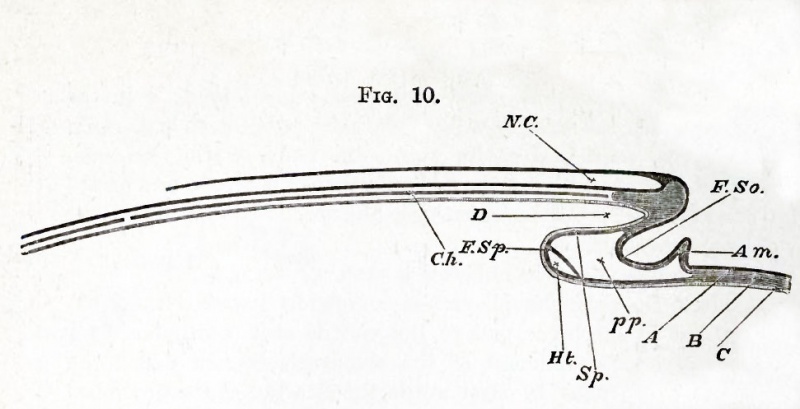
In a vertical longitudinal section carried through the middle line, we may recognize the following parts (Fig. 9, A, or on a larger scale Fig. 10, which also shews details which need not be considered now). Beginning at what will become the posterior extremity of the embryo (the left-hand side of the figure in each case), and following the surface of the blastoderm forwards (to the right in the figures), the level is maintained for some distance, and then there is a sudden descent, the blastoderm bending round and pursuing a precisely opposite direction to its previous one, running backwards instead of forwards, for some distance. It soon, however, turns round again, and once more running forward, with a gentle ascent, regains the original level. As seen in section, then, the blastoderm at this spot may be said to be folded up in the form of the letter "S". This fold we shall always speak of as the head-fold. In it we may recognize two limbs: an upper limb in which the curve is directed forwards, and its bay, opening backwards, is underneath the blastoderm, i.e. as we shall see, inside the embryo (Fig. 10. If) ; and an under limb in which the curve is directed backwards, and its bay, opening forwards, is above the blastoderm, i.e. outside the embryo. If an "S" like the above, made of some elastic material, were stretched laterally, the effect would be to make both limbs longer and proportionally narrower, and their bays, instead of being shallow cups, would become more tubular. Such a result is in part arrived at by the growth of the blastoderm; the upper limb of the "S" is continually growing forward (but, unlike the stretched elastic model, increases in all its dimensions at the same time), and the lower limb is as continually lengthening backwards; and thus both upper and lower bays become longer and longer. This we shall hereafter speak of as the travelling backwards of the head-fold.
Fig. 10. diagrammatic longitudinal section through the axis of an embryo.
- The section is supposed to be made at a time when the headfold has commenced but the tail-fold has not yet appeared. F. So. fold of the somatopleure. F. Sp. fold of the splanchnopleure.
- The line of reference F. So. is placed in the lower bay, outside the embryo. The line of D is placed in the upper bay inside the embryo ; this will remain as the alimentary canal. Both folds (F. So., F. Sp.) are parts of the head- fold, and are to be thought of as continually travelling onwards (to the left) as development proceeds. pp. space between somatopleure and splanchnopleure : pleuroperitoneal cavity.
Am. commencing (head) fold of the amnion. A fuller explanation is given under Fig. 29.
The two bays do not however both become tubular. The section we have been speaking of is supposed to be taken vertically along a line, which will afterwards become the axis of the embryo; and the lower bay of the 8 is a section of the crescentic groove mentioned above, in its middle or deepest part. On either side of the middle line the groove gradually becomes shallower. Hence in sections taken on either side of the middle line or axis of the embryo (above or below the plane of the figures), the groove would appear the less marked the farther the section from the middle line, and at a certain distance would disappear altogether. It must be remembered that the groove is at first crescent-shaped, with the concavity of the crescent turned towards what will be the hind end of the embryo (Fig. 22). As the whole head-fold is carried farther and farther back, the horns of the crescent are more and more drawn in towards the middle line, the groove becoming first semicircular, then horse-shoe-shaped. In other words, the head-fold, instead of being a simple fold running straight backwards, becomes a curved fold with a central portion in front running backwards, and two side portions running in towards the middle line. The effect of this is that the upper bay of the "S" (that within the embryo) gets closed in at the sides as well as in the front, and thus speedily becomes tubular. The under bay of the <3 (that outside the embryo) remains of course open at the sides as in front, and forms a sort of horse-shoe-shaped ditch surrounding the front end of the embryo.
We have dwelt thus at length on the formation of the head-fold, because, unless its characters are fairly grasped, much difficulty may be found in understanding many events in the history of the chick. The reader will perhaps find the matter easier to comprehend if he makes for himself a rough model, which he easily can do by spreading a cloth out flat to represent the blastoderm, placing one hand underneath it, to mark the axis of the embryo, and then tucking in the cloth from above under the tips of his fingers. The fingers, covered with the cloth and slightly projecting from the leve] of the rest of the cloth, will represent the head, in front of which will be the semicircular or horse-shoe-shaped groove of the head-fold.
At its first appearance the whole 8 may be spoken of as the head-fold, but later on it will be found convenient to restrict the name chiefly to the lower limb of the "S".
Some time after the appearance of the head-fold, an altogether similar but at first less conspicuous fold makes its appearance, at a point which will become the posterior end of the embryo. This fold, which travels forwards just as the head-fold travels backwards, is the tail-fold (Fig. 9, (7).
In addition, between the head- and the tail-fold two lateral folds appear, one on either side. These are simpler in character than either head-fold or tail-fold, inasmuch as they are nearly straight folds directed inwards towards the axis of the body (Fig. 8, F), and not complicated by being crescentic in form. Otherwise they are exactly similar, and in fact are formed by the continuations of the head- and tail-folds respectively.
As these several folds become more and more developed, the head-fold travelling backwards, the tailfold forwards, and the lateral folds inwards, they tend to unite in the middle point ; and thus give rise more and more distinctly to the appearance of a small tubular sac seated upon, and connected, by a continually-narrowing hollow stalk, with that larger sac which is formed by the extension of the rest of the blastoderm over the whole yolk.
The smaller sac we may call the " embryonic sac," the larger one " the yolk-sac." As incubation proceeds, the smaller sac (Fig. 9) gets larger and larger at the expense of the yolk-sac (the contents of the latter being gradually assimilated by nutritive processes into the tissues forming the growing walls of the former, not directly transferred from one cavity into the other). Within a day or two of the hatching of the chick, at a time when the yolk-sac is still of some considerable size, or at least has not yet dwindled away altogether, and the development of the embryonic sac is nearly complete, the yolk-sac (Fig. 9, N) is slipped into the body of the embryo, so that ultimately the embryonic sac alone remains.
The embryo, then, is formed by a folding-off of a portion of the blastoderm from the yolk-sac. The general outline of the embryo is due to the direction and shape of the several folds which share in its formation ; these, while preserving a nearly perfect bilateral symmetry, present marked differences at the two ends of the embryo. Hence from the very first there is no difficulty in distinguishing the end which will be the head from that which will be the tail.
In addition to this, the tubular sac of the embryo, while everywhere gradually acquiring thicker and thicker walls, undergoes at various points, through local activities of growth in the form of thickenings, ridges, buds or other processes, many modifications of the outline conferred upon it by the constituent folds. Thus bud-like processes start out from the trunk to form the rudiments of the limbs, and similar thickenings and ridges give rise to the jaws and other parts of the face. By the unequal development of these outgrowths the body of the chick is gradually moulded into its proper outward shape.
Were the changes which take place of this class only, the result would be a tubular sac of somewhat complicated outline, but still a simple tubular sac. Such a simple sac might perhaps be roughly taken to represent the body of many an invertebrate animal ; but the typical structure of a bird or other vertebrate animal is widely different. It may very briefly be described as follows.
First there is, above, a canal running lengthways along the body, in which are lodged the brain and spinal cord. Below this neural tube is an axis represented by the bodies of the vertebrae and their continuation forwards in the structures which form the base of the skull. Underneath this, again, is another tube closed in above by the axis, and on the sides and below by the body-walls. Enclosed in this second tube, and suspended from the axis, is a third tube, consisting of the alimentary canal with its appendages (liver, pancreas, lungs, &c., which are fundamentally mere diverticula from one simple canal). The cavity of the outer tube, which also contains the heart and other parts of the vascular system, is the general body cavity ; it consists of a thoracic or pleural, and an abdominal or peritoneal section ; these two parts are, however, from their mode of origin, portions of one and the same tube. Thus a transverse section of a vertebrate animal always shews the same fundamental structure : above a single tube, below a double tube, the latter consisting of one tube enclosed within another, the inner being the alimentary canal, the outer the general cavity of the body. Into such a triple tube the simple tubular embryonic sac of the chick is converted by a series of changes of a remarkable character.
The upper or neural tube is formed in the following way. At a very early period the upper layer of the blastoderm or epiblast in the region which will become the embryo, is raised up into two ridges or folds, which run parallel to each other at a short distance on either side of what will be the long axis of the embryo, and thus leave between them a shallow longitudinal groove (Fig. 9, B, also Figs. 21, m.c). As these ridges, which bear the name of medullary folds, increase in height they arch over towards each other, and eventually meet and coalesce in the middle line, thus converting the groove into a canal, which at the same time becomes closed at either end (Fig. 8, F. /, also Fig. 34. Me.}. The cavity so formed is the cavity of the neural tube, and eventually becomes the cerebro-spinal canal. Its walls are wholly formed of epiblast.
The lower double tube, that of the alimentary canal, and of the general cavity of the body, is formed in an entirely different way. It is, broadly speaking, the result of the junction and coalescence of the fundamental embryonic folds, the head-fold, tail-fold, and lateral folds ; in a certain sense the cavity of the body is the cavity of the tubular sac described in the last paragraph.
But it is obvious that a tubular sac formed by the folding-in of a single sheet of tissue, such as we have hitherto considered the blastoderm to be, must be a simple tubular sac possessing a single cavity only. The blastoderm however does not long remain a single sheet, but speedily becomes a double sheet of such a kind that, when folded in, it gives rise to a double tube.
Very early the blastoderm becomes thickened in the region of the embryo, the thickening being chiefly due to an increase in the middle layer or mesoblast, while at the same time it becomes split or cleft horizontally over the greater part of its extent into two leaves, an upper leaf and a lower leaf. In the neighbourhood of the axis of the body, beneath the neural tube, this cleavage is absent (Fig. 9, B ; also Figs. 24, 34), in fact, it begins at some little distance on either side of the axis and spreads thence into the periphery in all directions. It is along the mesoblast that the cleavage takes place, the upper part of the mesoblast uniting with epiblast to form the upper leaf, and the lower part with the hypoblast to form the lower leaf.
In the fundamental folds both leaves are involved, both leaves are folded downwards and inwards, both leaves tend to meet in the middle below ; but the lower leaf is folded in more rapidly, and thus diverges from the upper leaf, a space being gradually developed between them (Fig. 9). In course of time the several folds of the lower leaf meet and unite to form an inner tube quite independently of the upper leaf, whose own folds in turn meet and unite to form an outer tube separated from the inner one by an intervening space. The inner tube which from its mode of formation is clearly lined by hypoblast is the alimentary canal which is subsequently perforated at both ends to form the mouth and anus ; the walls of the outer tube are the walls of the body ; and the space between the two tubes is the general body or pleuroperitoneal cavity.
Hence the upper (or outer) leaf of the blastoderm, from its giving rise to the body-walls, is called the somatopleure (Soma, body, pleuron, side) ; the lower (or inner) leaf, from its form
ing the alimentary canal and its tributary viscera, the splanchnopleure (Splanchnon, viscus, pleuron, side).
This horizontal splitting of the blastoderm into a somatopleure and a splanchnopleure, which we shall hereafter speak of as the cleavage of the mesoblast, is not confined to the region of the embryo, but gradually extends over the whole of the yolk-sac. Hence in the later days of incubation the yolk-sac comes to have two distinct coats, an inner splanchnopleuric and an outer somatopleuric, separable from each other all over the sac. We have seen that, owing to the manner of its formation, the ' embryonic sac ' is connected with the ' yolk-sac ' by a continually narrowing hollow stalk ; but this stalk must, like the embryonic sac itself, be a double stalk, and consist of a smaller inner stalk within a larger outer one, Fig. 9, E } H. The folds of the splanchnopleure, as they tend to meet and unite in the middle line below, give rise to a continually narrowing hollow stalk of their own, a splanchnic stalk, by means of which the walls of the alimentary canal are continuous with the splanchnopleuric investment of the yolk-sac, and the interior of that canal is continuous with the cavity inside the yolk-sac. In the same way the folds of the somatopleure form a similar stalk of their own, a somatic stalk, by means of which the body- walls of the chick are continuous (for some time ; the continuity, as we shall see, being eventually broken by the development of the amnion) with the somatopleuric investment of the yolk-sac ; and the pleuroperitoneal cavity of the body of the chick is continuous with the narrow space between the two investments of the yolk-sac.
At a comparatively early period the canal of the splanchnic stalk becomes obliterated, so that the material of the yolk can no longer pass directly into the alimentary cavity, but has to find its way into the body of the chick by absorption through the bloodvessels. The somatic stalk, on the other hand, remains widely open for a much longer time ; but the somatic shell of the yolk-sac never undergoes that thickening which takes place in the somatic walls of the embryo itself; on the contrary, it remains thin and insignificant. When, accordingly, in the last days of incubation the greatly diminished yolk-sac with its splanchnic investment is withdrawn into the rapidly enlarging abdominal cavity of the embryo, the walls of the abdomen close in and unite, without any regard to the shrivelled, emptied somatopleuric investment of the yolk-sac, which is cast off as no longer of any use. (Fig. 9. Compare the series.)
The Amnion
Very closely connected with the cleavage of the mesoblast and the division into somatopleure and splanchnopleure, is the formation of the amnion, all mention of which was, for the sake of simplicity, purposely omitted in the description just given.
The amnion is a peculiar membrane enveloping the embryo, which takes its origin from certain folds of the somatopleure, and of the somatopleure only, in the following way.
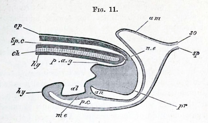
At a time when the cleavage of the mesoblast has somewhat advanced, there appears, a little way in front of the semilunar head-fold, a second fold (Fig. 22, also Fig. 9, (7.), running more or less parallel or rather concentric with the first, and not unlike it in general appearance, though differing widely from it in nature. In the head-fold the whole thickness of the blastoderm is involved ; in it both somatopleure and splanchnopleure (where they exist, i. e. where the mesoblast is cleft) take part. This second fold, on the contrary, is limited entirely to the somatopleure. Compare Figs. 9 and 10. In front of the head-fold, and therefore altogether in front of the body of the embryo, the somatopleure is a very thin membrane, consisting only of epiblast and a very thin layer of mesoblast ; and the fold we are speaking of is, in consequence, itself thin and delicate. Rising up as a semilunar fold with its concavity directed towards the embryo (Fig. 9, C, q/!), as it increases in height it is gradually drawn backwards over the developing head of the embryo. The fold thus covering the head is in due time accompanied by similar folds of the somatopleure starting at some little distance behind the tail, and at some little distance from the sides (Fig. 9, G, D, E, F, and Fig. 11 am.). In this way the embryo becomes surrounded by a series of folds of thin somatopleure, which form a continuous wall all round it. All are drawn gradually over the body of the embryo, and at last meet and completely coalesce (Fig. 9, H, 7), all traces of their junction being removed. Beneath these united folds there is therefore a cavity, within which the embryo lies (Fig. 9, H, ae). This cavity is the cavity of the amnion. The folds which we have been describing are those which form the amnion.
Fig. 11. Diagrammatic longitudinal section through the posterior end of an embryo bird, at the time of the formation of the allantois.
- ep. epiblast ; Sp.c. spinal canal ; ch. notochord ; n.e. neurenteric canal ; liy. hypoblast ; p.a.g. postanal gut ; pr. remains of primitive streak folded in on the ventral side ; al. allantois ; me. mesoblast; an. point where anus will be formed; p.c. perivisceral cavity ; am. amnion ; so. somatopleure ; sp. splanchnopleure.
Each fold, of course, necessarily consists of two limbs, both limbs consisting of epiblast and a very thin layer of mesoblast ; but in one limb the epiblast looks towards the embryo, while in the other it looks away from it. The space between the two limbs of the fold, as can easily be seen in Figs. 9 and 11, is really part of the space between the somatopleure and splanchnopleure ; it is therefore continuous with the general space, part of which afterwards becomes the pleuroperitoneal cavity of the body, shaded with dots in figure 9 and marked (p p). It is thus possible 'to pass from the cavity between the two limbs of each fold of the amnion into the cavity which surrounds the alimentary canal. When the several folds meet and coalesce together above the embryo, they unite in such a way that all their inner limbs go to form a continuous inner membrane or sac, and all their outer limbs a similarly continuous outer membrane or sac. The inner membrane thus built up forms a completely closed sac round the body of the embryo, and is called the amniotic sac, or amnion proper (Fig. 9, H, J, &c. a.), and the fluid which it afterwards contains is called the amniotic fluid, or liquor amnii. The space between the inner and outer sac, being formed by the united cavities of the several folds, is, from the mode of its formation, simply a part of the general cavity found everywhere between somatopleure and splanchnopleure. The outer sac over the embryo lies close under the vitelline membrane, while its periphery is gradually extended over the yolk as the somatopleuric investment of the yolk-sac described in the preceding paragraph. It constitutes the false amnion while the membrane of which it forms a part is frequently known as the serous membrane.
The Allantois
If the mode of origin of these two sacs (the inner or true amnion, and the outer or false amnion, as Baer called it) and their relations to the embryo be borne in mind, the reader will have no difficulty in understanding the course taken in its growth by an important organ, the allantois, of which we shall hereafter have to speak more in detail.
The allantois is essentially a diverticulum of the alimentary tract, into which it opens immediately in front of the anus. It at first (Fig. 11, al) forms a flattened sac projecting into the pleuroperitoneal cavity, the walls of the sac being formed of a layer of splanchnic mesoblast lined by hypoblast.
It grows forwards in the peritoneal cavity until it reaches the stalk connecting the embryo with the yolksac, and thence very rapidly pushes its way into the space between the true and false amniotic sacs (Fig. 9, G, K}. Curving over the embryo, it comes to lie above the embryo and the amnion proper, separated from the shell (and vitelline membrane) by nothing more than the thin false amnion. In this position it becomes highly vascular, and performs the functions of a respiratory organ. It is evident that though now placed quite outside the embryo, the space in which it lies is a continuation of that peritoneal cavity in which it took its origin.
It is only necessary to add, that the serous membrane, including the false amnion, either coalesces with the vitelline membrane, in contact with which it lies, or else replaces it ; and in the later days of incubation was called by the older embryologists the chorion a name however which we shall not adopt.
The Elements of Embryology - Volume 1 (1883)
The History of the Chick: Egg structure and incubation beginning | Summary whole incubation | First day | Second day - first half | Second day - second half | Third day | Fourth day | Fifth day | Sixth day to incubation end | Appendix
| Historic Disclaimer - information about historic embryology pages |
|---|
| Pages where the terms "Historic" (textbooks, papers, people, recommendations) appear on this site, and sections within pages where this disclaimer appears, indicate that the content and scientific understanding are specific to the time of publication. This means that while some scientific descriptions are still accurate, the terminology and interpretation of the developmental mechanisms reflect the understanding at the time of original publication and those of the preceding periods, these terms, interpretations and recommendations may not reflect our current scientific understanding. (More? Embryology History | Historic Embryology Papers) |
Glossary Links
- Glossary: A | B | C | D | E | F | G | H | I | J | K | L | M | N | O | P | Q | R | S | T | U | V | W | X | Y | Z | Numbers | Symbols | Term Link
Cite this page: Hill, M.A. (2024, April 28) Embryology Book - The Elements of Embryology - Chicken 2. Retrieved from https://embryology.med.unsw.edu.au/embryology/index.php/Book_-_The_Elements_of_Embryology_-_Chicken_2
- © Dr Mark Hill 2024, UNSW Embryology ISBN: 978 0 7334 2609 4 - UNSW CRICOS Provider Code No. 00098G

