Book - The Elements of Embryology - Chicken 1
| Embryology - 27 Apr 2024 |
|---|
| Google Translate - select your language from the list shown below (this will open a new external page) |
|
العربية | català | 中文 | 中國傳統的 | français | Deutsche | עִברִית | हिंदी | bahasa Indonesia | italiano | 日本語 | 한국어 | မြန်မာ | Pilipino | Polskie | português | ਪੰਜਾਬੀ ਦੇ | Română | русский | Español | Swahili | Svensk | ไทย | Türkçe | اردو | ייִדיש | Tiếng Việt These external translations are automated and may not be accurate. (More? About Translations) |
Foster M. Balfour FM. Sedgwick A. and Heape W. The Elements of Embryology (1883) Vol. 1. (2nd ed.). London: Macmillan and Co.
| Historic Disclaimer - information about historic embryology pages |
|---|
| Pages where the terms "Historic" (textbooks, papers, people, recommendations) appear on this site, and sections within pages where this disclaimer appears, indicate that the content and scientific understanding are specific to the time of publication. This means that while some scientific descriptions are still accurate, the terminology and interpretation of the developmental mechanisms reflect the understanding at the time of original publication and those of the preceding periods, these terms, interpretations and recommendations may not reflect our current scientific understanding. (More? Embryology History | Historic Embryology Papers) |
The structure of the hen's egg, and the changes which take place up to the beginning of incubation
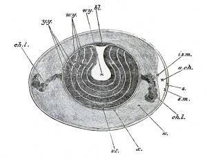
IN a hen's egg quite newly laid we meet with the following structures. Most external is the shell (Fig.1, s.) composed of an organic basis, impregnated with calcic salts. It is sufficiently porous to allow of the interchange of gases between its interior and the external air, and thus the chemical processes of respiration, feeble at first, but gradually increasing in intensity, are carried on during the whole period of incubation.
It is formed of two layers, both of which may contain pigment. The inner layer is by far the thickest, and is perforated by vertical canals which open freely on its inner aspect. Superficially these canals appear to be closed by the extremely thin outer layer. They are probably of some importance in facilitating the penetration of air through the shell.
Lining the shell, is the shell-membrane, which is double, being made up of two layers : an outer thicker (Fig. 1, s. m.), and an inner thinner one (i. s. m.). Both of these layers consist of several laminae of felted fibres of various sizes, intermediate in nature between connective and elastic fibres.
The white of the egg
Over the greater part of the egg the two layers of the shell-membrane remain permanently in close apposition ; but at the broad end they tend to separate, and thus to develope between them a space into which air finds its way. This air-chamber, as it is called, is not to be found in perfectly fresh eggs, but makes its appearance in eggs which have been kept for some time, whether incubated or not, and gradually increases in size, as the white of the egg shrinks in bulk from evaporation.
Immediately beneath the shell-membrane is the white of the egg or albumen (Fig. 1, w.), which is, chemically speaking, a mixture of various forms of proteid material, with fatty, extractive, and saline bodies. The outer part of the white, especially in eggs which are not perfectly fresh, is more fluid than that nearer the yolk.
Its average composition may be taken as 12*0 p. c. proteid matter, 1*5 p. c. fat and extractives, 5 p. c. saline matter, chiefly sodic and potassic chlorides, with phosphates and sulphates, 86*0 p. c. water.
The white of the egg when boiled shews in section alternate concentric layers of a transparent and of a finely granular opaque material. In the natural condition, the layers corresponding to these opaque layers are composed of more fluid albumen, while those corresponding to the transparent layers are less fluid, and consist of networks of fibres, containing fluid in their meshes. The innermost layer, however, immediately surrounding the yolk (Fig. 1, #.), is of the more fluid finely granular kind.
In eggs which have been hardened a spiral arrangement of the white may be observed, and it is possible to tear off laminae in a spiral direction from left to right, from the broad to the narrow end of the egg.
Two twisted cords called the chalazce (Fig. 1, ch. .), composed of coiled membranous layers of denser albumen, run from the two extremities of the egg to the opposite portions of the yolk. Their inner extremities expand and merge into a layer of denser albumen surrounding the fluid layer next the yolk. Their outer extremities are free, and do not quite reach the outer layer of the white. Thus they cannot serve to suspend the yolk, although they may help to keep it in position, by acting as elastic pads. The interior of each chalaza presents the appearance of a succession of opaque white knots ; hence the name chalazae (hailstones).
The yolk is enclosed in the vitelline membrane (Fig. 1, v. .), a transparent somewhat elastic membrane easily thrown into creases and wrinkles. It might almost be called structureless, but under a high power a fine fibrillation is visible, and a transverse section has a dotted or punctuated appearance ; it is probably therefore composed of fibrils. Its affinities are with elastic connective tissue.
The whole space within the vitelline membrane is occupied by the yolk. To the naked eye this appears tolerably uniform throughout, except at one particular point of its surface, at which may be seen, lying immediately under the vitelline membrane, a small white disc, about 4 mm. in diameter. This is the blastoderm, or cicatricula.
A tolerably typical cicatricula in a fecundated egg will shew an outer white rim of some little breadth, and within that a circular transparent area, in the centre of which, again, there is an opacity, varying in appearance, sometimes homogeneous, and sometimes dotted.
The disc is always found to be uppermost whatever be the position of the egg, provided there is no restraint to the rotation of the yolk. The explanation of this is to be sought for in the lighter specific gravity of that portion of the yolk which is in the neighbourhood of the disc, and the phenomenon is not in any way due to the action of the chalazae.
A section of the yolk of a hard-boiled egg will shew that it is not perfectly uniform throughout, but that there is a portion of it having the form of a flask, with a funnel-shaped neck, which, when the egg is boiled, does not become so solid as the rest of the yolk, but remains more or less fluid.
The expanded neck of this flask-shaped space is situated immediately underneath the disc, while its bulbous enlargement is about in the middle of the yolk. We shall return to it directly.
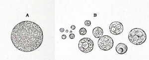
The great mass of the yolk is composed of what is known as the yellow yolk (Fig. 1, y. y.). This consists of spheres (Fig. 2, A.) of from 25/4 to lOOyu, 1 in diameter filled with numerous minute highly refractive granules ; these spheres are very delicate and easily destroyed by crushing. When boiled or otherwise hardened in situ, they assume a polyhedral form, from mutual pressure. The granules they contain seem to be of an albuminous nature, as they are insoluble in ether or alcohol.
Chemically speaking the yolk is characterized by the presence in large quantities of a proteid matter, having many affinities with globulin, and called vitellin. This exists in peculiar association with the remarkable body Lecithin. (Compare Hoppe-Seyler, Hdb. Phys. Chem. Anal.) Other fatty bodies, colouring matters, extractives (and, according to Dareste, starch in small quantities), &c. are also present. Miescher (Hoppe-Seyler, Chem. Untersuch. p. 502) states that a considerable quantity of nuclein may be obtained from the yolk, probably from the spherules of the white yolk.
The yellow yolk, thus forming the great mass of the entire yolk, is clothed externally by a thin layer of a different material, known as the white yolk, which at the edge of the blastoderm passes underneath the disc, and becoming thicker at this spot forms, as it were, a bed on which the blastoderm rests. Immediately under the middle of the blastoderm this bed of white yolk is connected, by a narrow neck, with a central mass of similar material, lying in the middle of the yolk (Fig. 1, w. y.). When boiled, or otherwise hardened, the white yolk does not become so solid as the yellow yolk ; hence the appearances to be seen in sections of the hardened yolk. The upper expanded extremity of this neck of white yolk is generally known as the "nucleus of Pander."
Concentric to the outer enveloping layer of white yolk there are within the yolk other inner layers of the same substance, which cause sections of the hardened yolk to appear to be composed of alternate concentric thicker laminae of darker (yellow) yolk, and thinner laminae of lighter (white) yolk (Fig. 1, w y ?/.).
The microscopical characters of the white yolk elements are very different from those of the yellow yolk. It is composed of vesicles (Fig. 2, J?.) for the most part smaller than those of the yellow yolk (4yi6 75^), with a highly refractive body, often as small as 1/-&, in the interior of each ; and also of larger spheres, each of which contains a number of spherules, similar to the smaller spheres.
Another feature of the white yolk, according to His, is that in the region of the blastoderm it contains numerous large vacuoles filled with fluid; they are sufficiently large to be seen with the naked eye, but do not seem to be present in the ripe ovarian ovum.
It is now necessary to return to the blastoderm. In this, as we have already said, the naked eye can distinguish an opaque white rim surrounding a more transparent central area, in the middle of which again is a white spot of variable appearance. In an unfecundated cicatricula the white disc is simply marked with a number of irregular clear spaces, there being no proper division into a transparent centre and an opaque rim.
The opaque rim is the commencement of what we shall henceforward speak of as the area opaca; the central transparent portion is in the same way the beginning of the area pellucida. In the part corresponding to the area opaca the blastoderm rests immediately on the white yolk ; underneath the area pellucida is a shallow space containing a nearly clear fluid, to the presence of which the central transparency seems to be due. The white spot in the middle of the area pellucida appears to be the nucleus of Pander shining through.
Vertical sections of the blastoderm shew that it is formed of two layers. The upper of these two layers is composed, see Fig. 3, ep, of a single row of cells, with their long axes arranged vertically, adhering together so as to form a distinct membrane, the edge of which rests upon the white yolk. After staining with silver nitrate, this membrane viewed from above shews a mosaic of uniform polygonal cells.
Each cell is composed of granular protoplasm filled with highly refractive globules ; and in each an oval nucleus may be distinguished. They are of a nearly uniform size (about 9 yu,) over the opaque and the pellucid areas.
The under layer (Fig. 3, I), is composed of cells which vary considerably in diameter; but even the smaller cells of this layer are larger than the cells of the upper layer. They are spherical, and so filled with granules and highly refractive globules, that a nucleus can rarely be seen in them : in the larger cells these globules are identical with the smaller white yolk spheres.
The cells of this layer do not form a distinct membrane like the cells of the upper layer, but lie as a somewhat irregular network of cells between the upper layer and the bed of white yolk on which the blastoderm rests. The lowest are generally the largest. The layer is thicker at the periphery than at the centre : and rests on a bed of white yolk, from which it is in parts separated by a more or less developed cavity, containing probably fluid yolk matter about to be absorbed. In the bed of white yolk nuclei are present, which are destined to become the nuclei of cells about to join the lower layer of the blastoderm. These nuclei are generally more numerous in the neighbourhood of the thickened periphery of the blastoderm than elsewhere. Amongst the lower layer cells are to be found peculiar large spherical bodies, which superficially resemble the larger cells around them, and have been called formative cells. Their real nature is still very doubtful, and though some are no doubt true cells, others are perhaps only nutritive masses of yolk.
The opacity of the peripheral part of the blastoderm is in a large measure due to the collection of the lower layer cells in this region, and the thickening, so caused, appears to be more pronounced for a small arc which subsequently constitutes the hinder border of the area pellucida.
Over nearly the whole of the blastoderm the upper layer rests on the under layer. At the circumference however the upper layer stretches for a short distance beyond the under layer, and here consequently rests directly on the white yolk.
To recapitulate : In the normal unincubated hen's egg we recognize the blastoderm, consisting of a complete upper layer of smaller nucleated granular cells and a more or less incomplete under layer of larger cells, filled with larger granules; in these lower cells nuclei are rarely visible. The thin flat disc so formed rests, at the uppermost part of the entire yolk, on a bed of white yolk, and a peripheral thickening of the lower layer causes the appearance in the blastodermic disc of an area opaca and an area pellucida. The great mass of the entire yolk consists of the so-called yellow yolk composed of granular spheres. The white yolk is composed of smaller spheres of peculiar structure, and exists, in small part, as a thin coating around, and as thin concentric laminae in the substance of the yellow yolk, but chiefly in the peculiar large spherical bodies, which superficially resemble the larger cells around them, and have been called formative cells. Their real nature is still very doubtful, and though some are no doubt true cells, others are perhaps only nutritive masses of yolk.
The opacity of the peripheral part of the blastoderm is in a large measure due to the collection of the lower layer cells in this region, and the thickening, so caused, appears to be more pronounced for a small arc which subsequently constitutes the hinder border of the area pellucida.
Over nearly the whole of the blastoderm the upper layer rests on the under layer. At the circumference however the upper layer stretches for a short distance beyond the under layer, and here consequently rests directly on the white yolk.
To recapitulate : In the normal unincubated hen's egg we recognize the blastoderm, consisting of a complete upper layer of smaller nucleated granular cells and a more or less incomplete under layer of larger cells, filled with larger granules; in these lower cells nuclei are rarely visible. The thin flat disc so formed rests, at the uppermost part of the entire yolk, on a bed of white yolk, and a peripheral thickening of the lower layer causes the appearance in the blastodermic disc of an area opaca and an area pellucida. The great mass of the entire yolk consists of the so-called yellow yolk composed of granular spheres. The white yolk is composed of smaller spheres of peculiar structure, and exists, in small part, as a thin coating around, and as thin concentric laminae in the substance of the yellow yolk, but chiefly in the form of a flask-shaped mass in the interior of the yolk, the upper somewhat expanded top of the neck of which forms the bed on which the blastoderm rests. The whole yolk is invested with the vitelline membrane, this again with the white ; and the whole is covered with two shell-membranes and a shell.
Such an egg has however undergone most important changes while still within the body of the hen; and in order to understand the nature of the structures which have just been described, it will be necessary to trace briefly the history of the egg from the stage when it exists as a so-called ovarian ovum in the ovary of a hen up to the time when it is laid.
In birds the left ovary alone is found in the adult ; and is attached by the mesovarium to the dorsal wall of the abdominal cavity, on the left side of the vertebral column. It consists of a mass of vascular stroma in which the ova are imbedded, is covered superficially by a layer of epithelium, continuous with the epithelial lining of the peritoneal cavity. The appearance of the ovary varies greatly according to the age of the individual. In the mature and sexually active females it is almost wholly formed of pedunculated and highly vascular capsules of various sizes, each containing a more or less developed ovum ; in the young animal however it is much more compact, owing to the absence of advanced ova.
If one of the largest capsules of the ovary of a hen which is laying regularly be opened, it will be found to contain a nearly spherical (or more correctly, ellipsoidal with but slightly unequal axes) yellow body enclosed in a delicate membrane. This is the ovarian ovum or egg.
Examined with care the ovum, which is tolerably uniform in appearance, will be found to be marked at one spot (generally facing the stalk of the capsule and forming the pole of the shorter axis of the ovum) by a small disc differing in appearance from the rest of the ovum. This disc which is known as the germinal disc or discus proligerus, consists of a lenticular mass of protoplasm (Fig. 4, c), imbedded in which is a globular or ellipsoidal body (Fig. 4, #), about SlO/,6 in diameter, called the germinal vesicle. This has a delicate wall, and its contents are clear and fluid in the fresh state, but become granular upon the addition of reagents.
Yolk of the egg
The rest of the ovum is known as the yolk. This consists of two elements, the white yolk- and the yellow yolk-spheres, which are distributed respectively very much in the same way as in the laid egg, the yellow yolk forming the main mass of the ovum, and the white yolk being gathered underneath and around the disc (Fig. 4, w. y\ and also forming a flask-shaped mass in the interior. The delicate membrane surrounding the whole is the vitelline membrane.
The youngest ova in the ovary of a fowl, in common with those of all other animals, present the characters of a simple cell. Such a cell is dia grammatically represented in Fig. 5.
It is seen to consist of a naked protoplasmic body containing in its interior a nucleus the germinal vesicle which in its turn envelopes a nucleolus constituting what is known as the germinal spot. Such young ova are enclosed in a capsule of epithelium, named the follicle or follicular membrane, and are irregularly scattered in the stroma of the ovary. The difference between such an immature ovum and the ripe ovum just described is very great, but throughout its growth the ovum retains the characters of a cell, so that the mature ovarian ovum, equally with the youngest ovum in the ovary, is a single cell. The most striking changes which takes place in the course of the maturation of the ovum concern the body of the cell rather than the germinal vesicle. As the body grows in size a number of granules make their appearance in its interior. These granules are formed by the inherent activity of the protoplasm, which is itself nourished, in a large measure at any rate, by the cells of the follicle. The outermost layer of the protoplasm remains free from these granules. As the ovum grows older the granules become larger, first of all in the centre, and subsequently at the periphery, and take the form of white yolk-spherules. The greater part of them become at a later stage converted into yellow yolk-spheres, while a portion of them, situated in the position of the white yolk of the ripe ovum, retain their original characters.
The germinal vesicle, which in the youngest ova is situated centrally or subcentrally, travels in the course of the growth of the ovum towards the periphery, and the protoplasm immediately surrounding it remains relatively free from yolk granules, and so constitutes the germinal disc. In the younger ova there is but a single germinal spot in the germinal vesicle, but as the ova enlarge several accessory germinal spots make their appearance, while in the ripe ovum it seems doubtful whether there is any longer a trace of a germinal spot.
The cells of the follicular epithelium are at first arranged in a single row, but at a later stage become two or more rows deep : they undergo however a nearly complete atrophy in the ripe ovum. Around the follicular epithelium, there is present a membrana propria, and in the later stages of the growth of the ovum this is in its turn embraced by a highly vascular connective-tissue capsule.
The youngest ova are, as has already been stated, quite naked. In ova of about 1*5 mm. the superficial layer of the ovum becomes converted into a radiately striated membrane called the zona radiata. At a later period a second membrane, placed between the zona radiata and the cells of the follicle, makes its appearance, but its mode of origin is still unknown. As the ovum approaches maturity the zona radiata disappears, and in the ripe ovum the second membrane, which has already been spoken of as the vitelline membrane, alone remains.
From what has just been stated it follows that in an egg which has been laid the yolk alone constitutes the true ovum. The white and the shell are in fact accessory structures formed during the passage of the ovum down the oviduct.
When the ovarian ovum is ripe and about to be discharged from the ovary, its capsule is clasped by the open infundibulum of the oviduct. The capsule then bursts, and the ovum escapes into the oviduct, its longer axis corresponding with the long axis of the oviduct, the germinal disc therefore being to one side.
In describing the changes which take place in the oviduct, it will be convenient, following the order previously adopted, to treat first of all of the formation of the accessory parts of the egg. These are secreted by the glandular walls of the oviduct. This organ therefore requires some description. It may be said to consist of four parts : 1st. The dilated infundibulum with an abdominal opening. 2nd. A long tubular portion the oviduct proper opening by a narrow neck or isthmus into the 3rd portion, which is much dilated, and has been called the uterus ; the 4th part is somewhat narrow, and leads from the uterus into the cloaca. The whole of the mucous membrane lining the oviduct is largely ciliated.
The accessory parts of the egg are entirely formed in the 2nd and 3rd portions. The layer of albumen which immediately surrounds the yolk is first deposited ; the chalazse are next formed. Their spiral character and the less distinctly marked spiral arrangement of the whole albumen is brought about by the motion of the egg along the spiral ridges into which the interior of the second or tubular portion of the oviduct is thrown. The spirals of the two chalazae are in different directions. This is probably produced by their peripheral ends remaining fixed while the yolk to which their central ends are attached is caused to rotate by the contractions of the oviduct. During the formation of the chalazse the rest of the albumen is also deposited ; and finally the shell-membrane is formed in the narrow neck of the 2nd portion, by the fibrillation of the most external layer of albumen. The egg passes through the 2nd portion in little more than 3 hours. In the 3rd portion the shell is formed. The mucous membrane of this part is raised into numerous flattened folds, like large villi, containing follicular glands. From these a thick white fluid is poured out, which soon forms a kind of covering to the egg, in which the inorganic particles are deposited. In this portion of the oviduct the egg remains from 12 to 18 hours, during which time the shell acquires its normal consistency. At the time of laying it is expelled from the uterus by violent muscular contractions, and passes with its narrow end downwards along the remainder of the oviduct, to reach the exterior.
Impregnation
This process occurs in the upper portion of the oviduct; the spermatozoa being found actively moving in a fluid which is there contained.
We have as yet, as far as the fowl is concerned, no direct observations concerning the changes preceding and following upon impregnation ; nor indeed concerning the actual nature of the act of impregnation.
In other types however these processes have been followed with considerable care, and the result has been to shew that prior to impregnation a division of the ovum takes place into two very unequal parts. The smaller of these parts is known as the polar body, and plays no further part in the development. In the course of the division of the ovum into these two parts the germinal vesicle also divides, and one part of it enters the polar body, while a portion remains in the larger segment which continues to be called the ovum, and is there known as the female pronudeus. Impregnation has been found to consist essentially in the entrance of a single spermatozoon into the ovum, followed by the fusion of the two. The spermatozoon itself is to be regarded as a cell, the head of which corresponds to the nucleus. When the spermatozoon enters the ovum the substance forming its tail becomes mingled with the protoplasm of the latter, but the head enlarges and constitutes a distinct body called the male pronudeus, which travels towards and finally fuses with the female pronucleus to constitute the nucleus of the impregnated ovum.
Segmentation
There follows upon the impregnation a remarkable process known as the segmentation. The process consists essentially in the division of the impregnated ovum by a series of successive segmentations into a number of cells, of which the whole of the cells of the future animal are the direct descendants. In the majority of instances this process results in the division of the whole ovum into cells ; but in cases of ova where there is a large amount of food yolk, only that part of the ovum in which the protoplasm is but slightly loaded with food material, and which we have already described as the germinal disc, becomes so divided. The remainder of the ovum constitutes a food reservoir for the use of the developing embryo and is known as the food yolk. The segmentation in such ova, of which that of the fowl is one of the best known examples, is described as being partial or meroblastic 1.
In order to understand the process of segmentation in the fowl's ovum it must be borne in mind that the germinal disc is not sharply separated from the remainder of the ovum, but that the two graduate insensibly into each other.
The segmentation commences in the lower part of the oviduct, shortly before the shell has begun to be formed.
(1 For a fuller account of the relation between holoblastic and meroblastic segmentation the reader is referred to the treatise on Comparative Embryology by Balfour, Vol. i. chapter iii.)
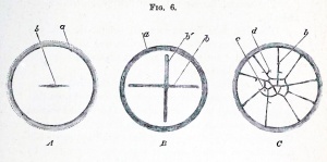
Viewed from above, a furrow is seen to make its appearance, running across the germinal disc, though not for the whole breadth, and dividing it into two halves (Fig. 6, A). This primary furrow is succeeded by a second at right angles to itself. The surface thus becomes divided into four segments or quadrants (Fig. 6, B).
The second furrow cuts the first somewhat excentrically.
The first four furrows do not extend through the whole thickness of the germinal disc, and the four segments marked out by them are not separated from the disc on their lower aspect.
Each of these is again bisected by radiating furrows, and thus the number of segments is increased from four to eight (it may be seven or nine). The central portion of each segment is then, by a cross furrow, cut off from the peripheral portion, giving rise to the appearance of a number of central smaller segments, surrounded by more external elongated segments (Fig. 6, (7).
The excentricity in the arrangement of the segments is moreover still preserved, the smaller segments being situated nearer one side of the germinal disc. The excentricity of the segmentation gives to the segmenting germinal disc a bilateral symmetry, but the relation between the axis of symmetry of the segmenting germinal disc and the long axis of the embryo is not known.
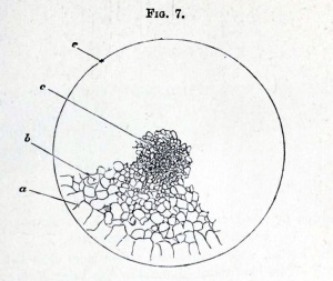
Division of the segments now proceeds rapidly by means of furrows running in various directions. And it is important to note that the central segments divide more rapidly than the peripheral, and consequently become at once smaller and more numerous (Fig. 7).
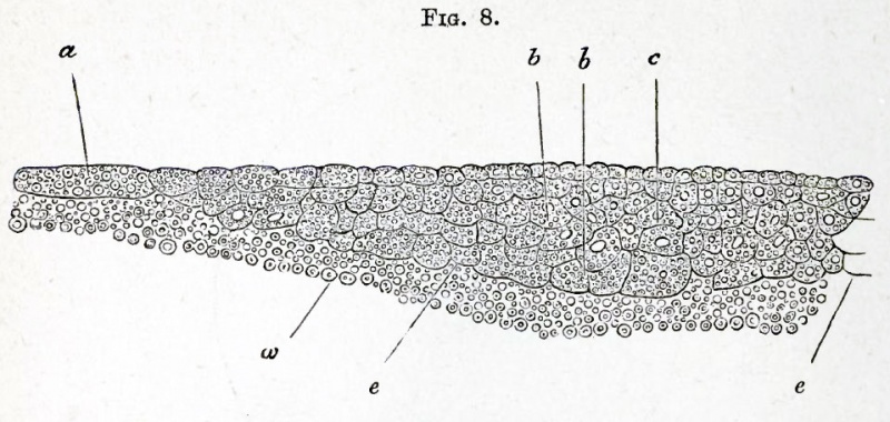
Meanwhile sections of the hardened blastoderm teach us that segmentation is not confined to the surface, but extends through the mass of the blastoderm ; they shew us moreover that division takes place by means of not only vertical, but also horizontal furrows, i. e. furrows parallel to the surface of the disc (Fig. 8).
At c in the centre of the disc the segmentation masses are very small and numerous. At b, nearer the edge, they are larger and fewer ; while those at the extreme margin a are largest and fewest of all. It will be noticed that the radiating furrows marking off the segments a do not reach to the extreme margin e of the disc.
The drawing is completed in one quadrant only ; it will of course be understood that the whole circle ought to be filled up in a precisely similar manner.
In this way, by repeated division or segmentation, the original germinal disc is cut up into a large number of small rounded masses of protoplasm, which are smallest in the centre, and increase in size towards the periphery. The segments lying uppermost are moreover smaller than those beneath, and thus the establishment of the two layers of the blastoderm is foreshadowed.
In the later stages of segmentation not only do the first-formed segments become further divided, but segmentation also extends into the remainder of the germinal disc.
The behaviour of the nucleus during the segmentation has not been satisfactorily followed, but there is, from the analogy of other forms, no doubt that in the formation of the first two segments the original nucleus, formed by the fusion of the male and female pronuclei, becomes divided, and that a fresh division of the nucleus takes place with the formation of each fresh segment. Nuclei make their appearance moreover in the part of the' ovum immediately below that in which the segmentation has already taken place ; these are in all probability also derived from the primitive nucleus. The substance round some of these nuclei rises up in the form of papillae, which are subsequently constricted off and set free as supplementary segmentation masses; while some of the nuclei remain and form the nuclei already spoken of as existing in the bed of white yolk below the blastoderm in the unincubated egg.
Between the segmented germinal disc, which we may now call the blastoderm, and the bed of white yolk on which it rests, a space containing fluid makes its appearance.
As development proceeds, segmentation reaches its limits in the centre, but continues at the periphery, and thus eventually the masses at the periphery become of the same size as those in the centre.
The distinction however between an upper and a lower layer becomes more and more obvious.
The masses of the upper layer arrange themselves, side by side, with their long axes vertical ; their nuclei become very distinct. In fact they form a membrane of columnar nucleated cells.
The masses of the lower layer, remaining larger than those of the upper layer, continue markedly granular and round, and form rather a close irregular network than a distinct membrane. Their nuclei are not readily visible.
At the time when the segmentation-spheres in the centre are smaller than those at the periphery, and those above are also smaller than those below, a few large spherical masses, probably containing each one of the nuclei already spoken of, arise by a process of segmentation from the bed of white yolk, and rest directly on the white yolk at the bottom of the shallow cavity below the mass of segmentation- spheres. They contain either numerous small spherules, or fine granules; the spherules precisely resembling the smaller spheres of white yolk. These loose spherical masses form the majority of the formative cells already spoken of.
Thus the original germinal disc of the ovarian ovum becomes, by the process of segmentation, converted into the blastoderm of the laid egg. with its upper layer of columnar nucleated cells, and its lower layer of irregularly disposed cells, accompanied by a few stray "formative " cells lying loose in the cavity below.
The Elements of Embryology - Volume 1 (1883)
The History of the Chick: Egg structure and incubation beginning | Summary whole incubation | First day | Second day - first half | Second day - second half | Third day | Fourth day | Fifth day | Sixth day to incubation end | Appendix
| Historic Disclaimer - information about historic embryology pages |
|---|
| Pages where the terms "Historic" (textbooks, papers, people, recommendations) appear on this site, and sections within pages where this disclaimer appears, indicate that the content and scientific understanding are specific to the time of publication. This means that while some scientific descriptions are still accurate, the terminology and interpretation of the developmental mechanisms reflect the understanding at the time of original publication and those of the preceding periods, these terms, interpretations and recommendations may not reflect our current scientific understanding. (More? Embryology History | Historic Embryology Papers) |
Glossary Links
- Glossary: A | B | C | D | E | F | G | H | I | J | K | L | M | N | O | P | Q | R | S | T | U | V | W | X | Y | Z | Numbers | Symbols | Term Link
Cite this page: Hill, M.A. (2024, April 27) Embryology Book - The Elements of Embryology - Chicken 1. Retrieved from https://embryology.med.unsw.edu.au/embryology/index.php/Book_-_The_Elements_of_Embryology_-_Chicken_1
- © Dr Mark Hill 2024, UNSW Embryology ISBN: 978 0 7334 2609 4 - UNSW CRICOS Provider Code No. 00098G


