Book - The Early Embryology of the Chick 4
| Embryology - 27 Feb 2026 |
|---|
| Google Translate - select your language from the list shown below (this will open a new external page) |
|
العربية | català | 中文 | 中國傳統的 | français | Deutsche | עִברִית | हिंदी | bahasa Indonesia | italiano | 日本語 | 한국어 | မြန်မာ | Pilipino | Polskie | português | ਪੰਜਾਬੀ ਦੇ | Română | русский | Español | Swahili | Svensk | ไทย | Türkçe | اردو | ייִדיש | Tiếng Việt These external translations are automated and may not be accurate. (More? About Translations) |
Patten BM. The Early Embryology of the Chick. (1920) Philadelphia: P. Blakiston's Son and Co.
| Online Editor |
|---|
| This historic 1920 paper by Bradley Patten described the understanding of chicken development. If like me you are interested in development, then these historic embryology textbooks are fascinating in the detail and interpretation of embryology at that given point in time. As with all historic texts, terminology and developmental descriptions may differ from our current understanding. There may also be errors in transcription or interpretation from the original text. Currently only the text has been made available online, figures will be added at a later date. My thanks to the Internet Archive for making the original scanned book available.
By the same author: Patten BM. Developmental defects at the foramen ovale. (1938) Am J Pathol. 14(2):135-162. PMID 19970381 Those interested in historic chicken development should also see the earlier text The Elements of Embryology (1883). Foster M. Balfour FM. Sedgwick A. and Heape W. The Elements of Embryology (1883) Vol. 1. (2nd ed.). London: Macmillan and Co.
Modern Notes |
| Historic Disclaimer - information about historic embryology pages |
|---|
| Pages where the terms "Historic" (textbooks, papers, people, recommendations) appear on this site, and sections within pages where this disclaimer appears, indicate that the content and scientific understanding are specific to the time of publication. This means that while some scientific descriptions are still accurate, the terminology and interpretation of the developmental mechanisms reflect the understanding at the time of original publication and those of the preceding periods, these terms, interpretations and recommendations may not reflect our current scientific understanding. (More? Embryology History | Historic Embryology Papers) |
The Establishment of the Entoderm
The Morula Stage
It should by no means be inferred that cell division ceases with the cleavage divisions. The end of the segmentation stage is not marked by even a retardation in the succession of mitoses. Segmentation is regarded as ending when the progress of development ceases to be indicated merely by increases in the number of cells, and begins to involve localized aggregation and differentiation of various groups of cells. Development progresses from phase to phase without abrupt change or interruption. The nomenclature and limitation of the various phases of development are largely arbitrary and the use of terms designating phases or stages of development should not be allowed to obscure the fact that the whole process is a continuous one.
In eggs without a large amount of yolk, segmentation results in the formation of a rounded, closely packed mass of blastomeres. This is known as a morula from its resemblance to the mulberry fruit which is in form much like the more familiar raspberry or blackberry. At the end of segmentation the chick embryo has arrived at a stage which corresponds with the morula stage of forms with less yolk. It consists of a discshaped mass of cells several strata in thickness, the blastoderm, lying closely applied to the yolk. In the center of the blastoderm the cells are smaller. and completely defined; at the periphery the cells are flattened, larger in surface extent, and are not walled off from the yolk beneath.
The Formation of the Blastula
The morula condition is of short duration. Almost as soon as it is established there begins a rearrangement of the cells presaging the formation of the blastula. A cavity is formed beneath the blastoderm by the detachment of its central cells from the underlying yolk while ^ the peripheral cells remain attached. The space thus established between the blastoderm and the yolk is termed the segmentation cavity (blastocoele). The marginal area of the blastoderm in which the cells remain undetached from the yolk and closely adherent to it, is called the zone of junction. With the establishment of the blastocoele the embryo is said to have progressed from the morula to the blastula stage.
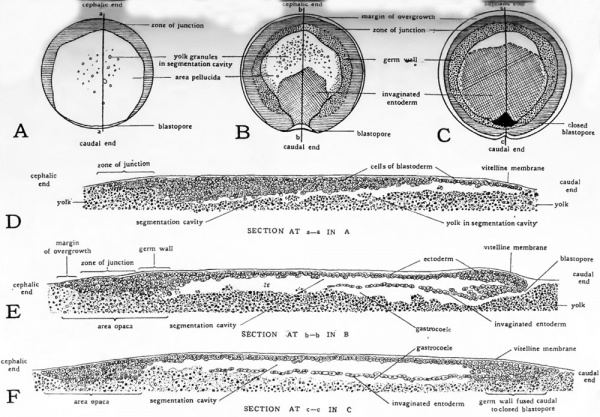
Figure 7, D, shows the conditions seen on sectioning the blastula of a bird. Only the blastoderm and the immediately underlying yolk are included in the diagram. At this magnification the complete yolk must be imagined as about three feet in diameter. The structure of the bird embryo in these stages may be brought in line with the morula and blastula stages of forms having little yolk if the full significance of the great yolk mass is appreciated. Instead of being free to aggregate first into a solid sphere of cells (morula) and then into a hollow sphere of cells (blastula), as takes place in forms with httle yolk, the blastomeres in the bird embryo are forced to grow on the surface of a large yolk sphere. Under such mechanical conditions the blastomeres are forced to become arranged in a disc-shaped mass on the surface of the yolk. If one imagines the yolk of the bird morula removed, and the disc of cells left free to assume the spherical shape dictated by surface tension its comparability with the morula in a form having little yolk becomes apparent.
The process of blastulation also is modified by the presence of a large amount of yolk. There can be no simple hollow sphere formation by rearrangement of the cells if the great bulk of the morula is inert yolk. But the cells of the central region of the blastoderm are nevertheless separated from the yolk to form a small blastocoele. The yolk constitutes the floor of the blastocoele and at the same time by reason of its great mass nearly obliterates it. If we imagine the yolk removed from the blastula and the edges of the blastoderm pulled together the chick blastula approaches the form of the blastula in embryos with little yolk.
The Effect of Yolk on Gastrulation
The process of gastrulation begins as soon as blastulation is accomplished. Gastrulation as it occurs in birds is not difiicult to understand if one grasps its fundamental similarity to the corresponding process in forms with scanty yolk. In Amphioxus, gastrulation is an inpocketing of the blastula (Fig. 6). A double layered cup is formed from a single layered hollow sphere much as one might push in a hollow rubber ball with the thumb. The new cavity in the double walled cup is termed the gastrocoele. The opening from the outside into the gastrocoele is called the blastopore.
Fig. 6. Schematic diagrams to show the effect of yolk on gastrulation.
- Abbreviations: blc, blastocoele; bid., blastoderm; blp., blastopore; ect., ectoderm; ent., entoderm; mit., cell undergoing mitosis; yk., yolk; yk.g., yolk granules; yk.p., yolk plug.
In gastrulation the single cell layer of the blastula is doubled upon itself to form two layers. The outer cell layer is known as the ectoderm and the inner layer as the entoderm. These layers differ from each other in their positional relationship to the embryo and to the surrounding environment. Each has different functional potentialities and each will in the course of development give rise to quite different types of structures and organs. It is the importance of their later history rather than any complexity or veiled significance about the way in which they arise that attaches such importance in embryology to the estabhshment of these two layers.
In the gastrulation of Amphibian embryos (Fig. 6) the yolk forces the invagination of the blastoderm toward the animal pole, but the inpocketing takes place into the blastocoele and the interrelationships of ectoderm, entoderm, and gastrocoele are established in fundamentally the same way as in Amphioxus. Gastrulation in birds is greatly modified by the large amount of yolk present (Fig. 6). Infolding must be effected in a disc of cells resting like a cap on a large yolk sphere. The smallness of the blastocoele sharply restricts the space into which the invagination can grow. Instead of arising as a relatively large circular opening the blastopore appears as a crescentic slit at the margin of the blastoderm. The crescentic blastopore may be regarded as a potentially circular opening which has been flattened as it develops between the growing disc of cells and the unyielding yolk which under lies them. The invaginated pocket of entoderm which grows in from this compressed blastopore is also flattened, conforming to the restrictions of the shape and size of the blastocoele. Moreover the floor of the invagination is represented only by a few widely scattered cells lying upon the yolk. It is as if the lower layer in its ingrowth was impeded and broken up by the yolk. The scattered cells representing the floor of the invagination soon disappear and the yolk itself comes to constitute the floor of the gastrocoele. Notwithstanding the great displacement of the blastopore and the gastrular invagination toward the animal pole and the restricted size and incomplete floor of the gastrocoele, the cell layers and the cavity established can be homologized with the corresponding features in forms where the course of development has not been so extensively modified by yolk.
A comparative review of the diagrams of Figure 6 will afford a general understanding of the infolding process of gastrulation. These diagrams aim to convey merely the scheme of the process. They are therefore simplified and emphasize the similarities of gastrulation in forms with widely varying amounts of yolk, rather than the details of the process in any one form. With this general groundwork we may now profitably return to the blastula stage and consider in somewhat more detail the process of gastrulation as it occurs in birds.
Gastrulation in Birds
We have already estabUshed the blastula as a disc of cells lying on the yolk but separated from it centrally by a flattened blastocoele or segmentation cavity. The peripheral part of the blastoderm where the marginal cells lie unseparated from the yolk has been termed the zone of junction (Fig. 7, D). This part of the blastoderm is also called the area opaca because in preparations made by removing the blastoderm from the yolk surface, yolk adheres to it and renders it more opaque. This opacity is especially apparent when a preparation is viewed under the microscope by transmitted light. The central area of the blastoderm, because it is separated from the yolk by the segmentation cavity, does not bring a mass of adherent yolk with it when the blastoderm is removed.
.It is for this reason translucent and is called the area pellucida. The area opaca later becomes differentiated so that three more or less distinct zones may be distinguished: (i) a peripheral zone known as the margin of overgrowth where rapid proliferation has pushed the cells out over the yolk without their becoming adherent to it; (2) an intermediate zone known as the zone of junction in which the deep-lying cells do not have complete cell boundaries but constitute a syncytium blending without definite boundary into the superficial layer of white yolk and adhering to it by means of penetrating strands of cytoplasm; (3) an inner zone known as the germ wall made up of cells derived from the inner border of the zone of junction which have acquired definite boundaries and become more or less free from the yolk. The cells of the germ wall usually contain numerous small yolk granules which were enmeshed in their cytoplasm when they were, as cells of the zone of junction, unseparated from the yolk (Fig. 7, J5, E). The inner margin of the germ wall marks the transition from area opaca to area pellucida.
Fig. 7. Diagrams to show various stages in the gastrulation of a bird. (After Patterson's figures for the pigeon.)
The changes in the blastula which indicate the approach of gastrulation are, first, a thinning of the blastoderm at its caudal margin and, second, freeing of the blastoderm from the yolk in the same region (Fig. 7, Z)). The separation of the blastoderm from the yolk is evidenced in surface views by a crescentic gap in the posterior quadrant of the zone of junction (Fig. 7, A). This region where the blastoderm is thin and free from the yolk marks the position of the blastopore.
Gastrulation begins with the undertucking of the cells at the free margin of the blastoderm. Figure 7, 5, is a diagrammatic surface view of the blastoderm represented as a transparent object. The position and the extent of the invaginated entoderm seen .through the overlying ectoderm are indicated by the cross hatched area. The appearance of the blastopore locates the caudal region of the future embryo and permits the definition of its longitudinal axis. This axis is indicated by the line h-h on Figure 7, J5. A diagram of a section cut in the longitudinal axis and passing through the blastopore of an embryo of this stage is shown in Figure 7, E. The invaginated cells which constitute the entoderm form a layer extending cephalad from the thickened lip of the blastopore. The yolk forms the floor of the gastrocoele. Figure 7, C, is a diagrammatic surface- view of a later stage in the same process. The extent of the entoderm is marked by cross-hatching as in the diagram of the previous stage. The undertucking of the cells at the blastopore has ceased by this time, and as indicated in Figure .7, C. by the black area, and in Figure 7, by the solid mass of cells seen in section, the blastopore has become closed.
During the entire time that the process of gastrulation has been in progress there has been constant cell proliferation going on in the blastoderm as a whole. The growth of the blastoderm has been evidenced especially by increase in its surface extent which has resulted in a general spreading of its peripheral margins over the yolk. This extension has taken place uniformly at all parts of the margin except in the posterior quadrant where the blastopore is located. Here the cells proliferated, instead of spreading out over the yolk have turned in at the lip of the blastopore to form the invaginated entoderm. This particular part of the margin of the blastoderm, having contributed the cells formed in its growth to the entoderm which grows back toward the center of the blastoderm, takes no part in the general peripheral expansion. As a result the blastopore region is, as it were, left behind and the rapidly extending margin of the blastoderm on either side sweeps around and encloses it. The blastopore at the time of its closure thus comes to lie within the recompleted circle of the germ wall (Fig. 7, C).
The Early Embryology of the Chick: Introduction | Gametes and Fertilization | Segmentation | Entoderm | Primitive Streak and Mesoderm | Primitive Streak to Somites | 24 Hours | 24 to 33 Hours | 33 to 39 Hours | 40 to 50 Hours | Extra-embryonic Membranes | 50 to 55 Hours | Day 3 to 4 | References | Figures | Site links: Embryology History | Chicken Development
| Historic Disclaimer - information about historic embryology pages |
|---|
| Pages where the terms "Historic" (textbooks, papers, people, recommendations) appear on this site, and sections within pages where this disclaimer appears, indicate that the content and scientific understanding are specific to the time of publication. This means that while some scientific descriptions are still accurate, the terminology and interpretation of the developmental mechanisms reflect the understanding at the time of original publication and those of the preceding periods, these terms, interpretations and recommendations may not reflect our current scientific understanding. (More? Embryology History | Historic Embryology Papers) |
Glossary Links
- Glossary: A | B | C | D | E | F | G | H | I | J | K | L | M | N | O | P | Q | R | S | T | U | V | W | X | Y | Z | Numbers | Symbols | Term Link
Cite this page: Hill, M.A. (2026, February 27) Embryology Book - The Early Embryology of the Chick 4. Retrieved from https://embryology.med.unsw.edu.au/embryology/index.php/Book_-_The_Early_Embryology_of_the_Chick_4
- © Dr Mark Hill 2026, UNSW Embryology ISBN: 978 0 7334 2609 4 - UNSW CRICOS Provider Code No. 00098G


