Book - The Early Embryology of the Chick 2
| Embryology - 27 Feb 2026 |
|---|
| Google Translate - select your language from the list shown below (this will open a new external page) |
|
العربية | català | 中文 | 中國傳統的 | français | Deutsche | עִברִית | हिंदी | bahasa Indonesia | italiano | 日本語 | 한국어 | မြန်မာ | Pilipino | Polskie | português | ਪੰਜਾਬੀ ਦੇ | Română | русский | Español | Swahili | Svensk | ไทย | Türkçe | اردو | ייִדיש | Tiếng Việt These external translations are automated and may not be accurate. (More? About Translations) |
Patten BM. The Early Embryology of the Chick. (1920) Philadelphia: P. Blakiston's Son and Co.
| Online Editor |
|---|
| This historic 1920 paper by Bradley Patten described the understanding of chicken development. If like me you are interested in development, then these historic embryology textbooks are fascinating in the detail and interpretation of embryology at that given point in time. As with all historic texts, terminology and developmental descriptions may differ from our current understanding. There may also be errors in transcription or interpretation from the original text. Currently only the text has been made available online, figures will be added at a later date. My thanks to the Internet Archive for making the original scanned book available.
By the same author: Patten BM. Developmental defects at the foramen ovale. (1938) Am J Pathol. 14(2):135-162. PMID 19970381 Those interested in historic chicken development should also see the earlier text The Elements of Embryology (1883). Foster M. Balfour FM. Sedgwick A. and Heape W. The Elements of Embryology (1883) Vol. 1. (2nd ed.). London: Macmillan and Co.
Modern Notes |
| Historic Disclaimer - information about historic embryology pages |
|---|
| Pages where the terms "Historic" (textbooks, papers, people, recommendations) appear on this site, and sections within pages where this disclaimer appears, indicate that the content and scientific understanding are specific to the time of publication. This means that while some scientific descriptions are still accurate, the terminology and interpretation of the developmental mechanisms reflect the understanding at the time of original publication and those of the preceding periods, these terms, interpretations and recommendations may not reflect our current scientific understanding. (More? Embryology History | Historic Embryology Papers) |
The Gametes and Fertilization
The Ovarian Ovum
The formation of the ovum, the phenomena of fertihzation, and the stages of development occurring prior to the laying of the egg have been more completely worked out in the pigeon than in the hen. The observations which have been carried out on the hen's egg indicate, as might be expected from the near relationship of the pigeon and the hen, that the processes in the two forms are closely comparable. The following account which is based chiefly on observations made on the pigeon's egg may, therefore, be taken to apply equally well in all essentials to the hen's egg.
The part of the egg commonly known as the "yolk" is a single cell, the female sex cell or ovum. Its great size as compared with other cells is due to the food material it contains. While the egg cell is still in the ovary, material which is later used by the embryo as food is deposited in its cytoplasm. This deposit which is known as deutoplasm consists of a viscid fluid in which are suspended granules and globules of various sizes. As the deutoplasm increases in amount the nucleus and the cytoplasm are forced toward the surface so that eventually the deutoplasm comes to occupy nearly the entire cell. This abundance of deutoplasm accumulated in the ovum furnishes a readily assimilable food supply, which makes possible the extreemly rapid development of the chick embryo.
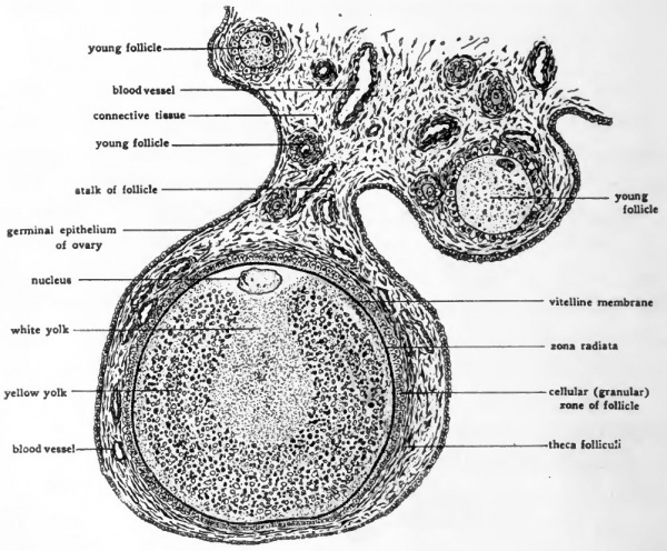
A section of the hen's ovary passing through a nearly mature ovum (Fig. 1) shows the ovum and the tissues which surround it projecting from the ovary but connected to it by a constricted stalk of ovarian tissue. The protuberance containing the ovum is known as a follicle. The bulk of the ovum itself is made up of the yolk. Except in the neighborhood of the nucleus the active cytoplasm is but a thin film enveloping the yolk. About the nucleus a considerable mass of cytoplasm is aggregated. The region of the ovum containing the nucleus and the bulk of the active cytoplasm is known as the animal pole because this subsequently becomes the site of greatest protoplasmic activity. The region opposite the animal pole is called the vegetative pole because while material for growth is drawn from this region it remains itself relatively inactive.
Fig. 1. Diagram showing the structure of a bird ovum still in the ovary. (Modified from Lillie, after Patterson.) The section shows a follicle containing a nearly mature ovum, together with a small area of the adjacent overian tissue.
Enclosing the ovum is a thin non-cellular membrane, the vitelline membrane, which is a secretory product of the cytoplasm of the ovum. Outside the vitelline membrane and very difficult to differentiate from it, is another, secreted membrane the zona radiata, so called because of its delicate radial striations. Immediately peripheral to the zona radiata is an investment of small polygonal cells, the cellular or "granular" zone of the follicle, which is in turn enclosed in a highly vascular coat of connective tissue, the theca folliculi. The nutriment for the growing ovum is supplied by the mother from the products ot her digested food. It is brought in through the blood vessels of the theca, absorbed by the foliicular cells and transferred by them to the ovum. Within the ovum this material is elaborated into deutoplasm.
Maturation, Ovulation and Fertilization
When the full allotment of deutoplasm has accumulated in the ovum the nucleus undergoes its first maturation division. Maturation is a process occurring before fertilization, in which there is an equal mitotic division of the nucleus of the ovum but a markedly unequal division of the cytoplasm and its contents. The result of this division is the formation of one very large cell containing the entire dower of deutoplasm and one very small cell containing practically no deutoplasm. This small cell is called a polar body because it is budded off at the animal pole of the ovum. Since this unequal division of the ovum typically occurs twice we speak of the first and second maturation divisions and of the first and second polar bodies.
In one of these maturation divisions the chromosomes do not split at the metaphase stage as happens in ordinary mitoses. Instead, half of the original number of chromosomes migrate bodily to each pole of the spindle, with the result that each daughter nucleus receives but half the number of chromosomes normal for the somatic cells of the species. Such a modified mitotic division is known as a reduction division. After the maturation divisions, one of which is a reduction division, the nucleus of the ovum now ready for fertilization,- is called the female pronucleus.
Although maturation in the male sex cells differs in some respects from the maturation of the ovum, there also, a reduction division occurs. The result is that the nucleus of each matured cell contains but half the species number of chromosomes. When in the process of fertilization the nucleus of the male cell unites with the female pronucleus the full species number of chromosomes is restored.
At about the time of the first maturation division the follicle ruptures, and the liberated ovum passes into the oviduct. If insemination has taken place meanwhile, the spermatozoa (Fig. 2) make their way along the oviduct where for several days they may remain alive and capable of performing their function of fertilization. Penetration of the ovum by spermatozoa takes place in the region of the oviduct near the ovary, before the albumen and shell have been added to the ovum. Coincidently the second polar body is extruded. Although in birds normally several spermatozoa penetrate the ovum, only a single one unites with the female pronucleus. The fusion of the male and female pronuclei in fertilization initiates the development of the embryo and the cleavage divisions are begun while the ovum is passing through the oviduct toward the cloaca and receiving meanwhile its accessory coverings.
The Formation of the Accessory Coverings of the Ovum
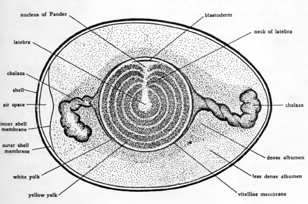
The albumen, the shell membrane, and the shell are non-cellular investments secreted about the ovum by the cells hning the oviduct. In the part of the oviduct adjacent to the ovary a mass of stringy albuminous material is produced. This adheres closely to the vitelline membrane and projects beyond it in two masses extending in either direction along the oviduct. Due to the spirally arranged folds in the walls of the oviduct, the egg as it moves toward the cloaca is rotated. This rotation twists the adherent albumen into the form of spiral strands projecting at either end of the yolk, known as the chalazae (Fig. 3). Additional albumen, which has been secreted abundantly in advance of the ovum by the glandular lining of the oviduct, is caught in the chalazae and during the further descent of the ovum is wrapped about it in concentric layers. These lamellae of albumen may be easily demonstrated in an egg which has had the albumen coagulated by boiling. The albumen secreting region of the oviduct constitutes about one-half of its entire length.
The shell membranes which consist of sheets of matted organic fibers are added farther along in the oviduct. The shell is secreted as the egg is passing through the shell gland portion of the oviduct. The entire passage of the ovum from the time of its discharge from the ovary to the time when it is ready for laying has been estimated to occupy about 22 hours If the completely formed egg reaches the cloacal end of th oviduct during the middle of the day it is usually laid at once otherwise it is likely to be retained until the following day. This over night retention of the egg is one of the factors which accounts for the variability in the stage of development reached at the time of laying.
The Structure of the Egg at the Time of Laying
The arrangement of structures in the egg at the time of laying is shown in Figure 3. Most of the gross relationships are already familiar because they appear so clearly in eggs which have been boiled. If a newly laid egg is allowed to float free in water until it comes to rest and is then opened by cutting away the part of the shell which lies uppermost, a circular whitish area will be seen to lie atop the yolk. In eggs which have been fertilized this area is somewhat different in appearance and noticeably larger than it is in unfertilized eggs. The differences are due to the development which has taken place in fertilized eggs during their passage through the oviduct. The aggregation of cells which in fertilized eggs lies in this area is known as the blastoderm. The structure of the blastoderm and the manner in which it grows will be taken up in the next chapter.
Fig. 3. Diagram of the hen's egg in longitudinal section. (After Lillie.) The relations of the various parts of the egg at the time of laying are indicated schematically.
Close examination of the yolk will show that it is not uniform throughout either in color or in texture. Two kinds of yolk can be differentiated, white yolk, and yellow yolk. Aside from the difference in color visible to the unaided eye, microscopical examination will show that there are differences in the granules and globules of the two types of yolk, those in the white yolk being in general smaller and less uniform in appearance. The principal accumulation of white yolk lies in a central flaskshaped area, the latebra, which extends toward the blastoderm and flares out under it into a mass known as the nucleus of Pander. In addition to the latebra and the nucleus of Pander there are thin concentric layers of white yolk between which lie much thicker layers of yellow yolk. The concentric layers of white and yellow yolk are said to indicate the daily accumulation of deutoplasm during the final stages in the formation of the egg. The outermost yolk immediately under the vitelline membrane is always of the white variety.
The albumen, except for the chalazae, is nearly homogeneous in appearance, but near the yolk it is somewhat more dense than it is peripherally. The chalazae serve to suspend the yolk in the albumen.
The two layers of shell membrane lie in contact everywhere except at the large end of the egg where the inner and outer membranes are separated to form an air chamber. This space is stated (Kaupp) to appear only after the egg has been laid and cooled from the body temperature of the hen (about io6°F.) to the ordinary temperatures. In eggs which have been kept for any length of time the air space increases in size due to evaporation of part of the water content of the egg. This fact is taken advantage of in the familiar method of testing the freshness of eggs by floating them."
The egg shell is composed largely of calcareous salts. These salts are derived from the food of the mother and if lime containing substances are not furnished in her diet the shell is defectively formed or even altogether wanting. The shell is porous allowing the embryo to carry on exchange of gases with the outside air by means of • specialized vascular membranes arising in connection with the embryo but lying outside it, directly beneath the shell.
Incubation
When an egg has been laid, development ceases unless the temperature of the egg is kept nearly up to the body temperature of the mother. Cooling of the egg does not, however, result in the death of the embryo. It may resume its development if it is brooded by the hen or artificially incubated even after the egg has been kept for many days at ordinary temperatures.
The normal incubation temperature is that at which the egg is maintained by the body heat from the brood-hen. This is somewhat below the blood heat of the hen (106oF.). When an egg is allowed to remain undisturbed the yolk rotates so that the developing embryo lies uppermost. Its position is then such that it gets the full benefit of the warmth of the mother.
In incubating eggs artificially the incubators are usually regulated for a heat of 100o-101oF. (37o-38oC.). At this temperature the chick is ready for hatching on the twenty-first day. Development will go on at considerably lower temperatures but its rate is retarded in proportion to the lowering of the temperature. Below about 21 degrees Centigrade development ceases altogether.
In incubating eggs which have been cooled after laying for some particular stage of the embryo which it is desired to secure, three or four hours are ordinarily allowed for the egg to become warmed to the point at which development begins again. For example if an embryo of 24-hours incubation age is desired the egg should be allowed to remain in the incubator about 27 hours. Even with allowance* made for the warming of the egg and with exact regulation of the temperature of the incubator, the stage of development attained in a given incubation time will vary widely in different eggs. The factor of individual variability which must always be reckoned with in developmental processes, undoubtedly accounts for some of the variation. The different time occupied by different eggs in traversing the oviduct, the over-night retention of eggs not ready for laying till toward sundown, and especially the varying time different eggs have been brooded before being removed from the nest, account for further variations. The designation of the age of chicks in hours of incubation is, therefore, not exact, but merely a convenient approximation of the average condition reached in that incubation time.
- Next: Segmentation
The Early Embryology of the Chick: Introduction | Gametes and Fertilization | Segmentation | Entoderm | Primitive Streak and Mesoderm | Primitive Streak to Somites | 24 Hours | 24 to 33 Hours | 33 to 39 Hours | 40 to 50 Hours | Extra-embryonic Membranes | 50 to 55 Hours | Day 3 to 4 | References | Figures | Site links: Embryology History | Chicken Development
| Historic Disclaimer - information about historic embryology pages |
|---|
| Pages where the terms "Historic" (textbooks, papers, people, recommendations) appear on this site, and sections within pages where this disclaimer appears, indicate that the content and scientific understanding are specific to the time of publication. This means that while some scientific descriptions are still accurate, the terminology and interpretation of the developmental mechanisms reflect the understanding at the time of original publication and those of the preceding periods, these terms, interpretations and recommendations may not reflect our current scientific understanding. (More? Embryology History | Historic Embryology Papers) |
Glossary Links
- Glossary: A | B | C | D | E | F | G | H | I | J | K | L | M | N | O | P | Q | R | S | T | U | V | W | X | Y | Z | Numbers | Symbols | Term Link
Cite this page: Hill, M.A. (2026, February 27) Embryology Book - The Early Embryology of the Chick 2. Retrieved from https://embryology.med.unsw.edu.au/embryology/index.php/Book_-_The_Early_Embryology_of_the_Chick_2
- © Dr Mark Hill 2026, UNSW Embryology ISBN: 978 0 7334 2609 4 - UNSW CRICOS Provider Code No. 00098G


