Book - The Early Embryology of the Chick 10
| Embryology - 30 Apr 2024 |
|---|
| Google Translate - select your language from the list shown below (this will open a new external page) |
|
العربية | català | 中文 | 中國傳統的 | français | Deutsche | עִברִית | हिंदी | bahasa Indonesia | italiano | 日本語 | 한국어 | မြန်မာ | Pilipino | Polskie | português | ਪੰਜਾਬੀ ਦੇ | Română | русский | Español | Swahili | Svensk | ไทย | Türkçe | اردو | ייִדיש | Tiếng Việt These external translations are automated and may not be accurate. (More? About Translations) |
Patten BM. The Early Embryology of the Chick. (1920) Philadelphia: P. Blakiston's Son and Co.
| Online Editor |
|---|
| This historic 1920 paper by Bradley Patten described the understanding of chicken development. If like me you are interested in development, then these historic embryology textbooks are fascinating in the detail and interpretation of embryology at that given point in time. As with all historic texts, terminology and developmental descriptions may differ from our current understanding. There may also be errors in transcription or interpretation from the original text. Currently only the text has been made available online, figures will be added at a later date. My thanks to the Internet Archive for making the original scanned book available.
By the same author: Patten BM. Developmental defects at the foramen ovale. (1938) Am J Pathol. 14(2):135-162. PMID 19970381 Those interested in historic chicken development should also see the earlier text The Elements of Embryology (1883). Foster M. Balfour FM. Sedgwick A. and Heape W. The Elements of Embryology (1883) Vol. 1. (2nd ed.). London: Macmillan and Co.
Modern Notes |
| Historic Disclaimer - information about historic embryology pages |
|---|
| Pages where the terms "Historic" (textbooks, papers, people, recommendations) appear on this site, and sections within pages where this disclaimer appears, indicate that the content and scientific understanding are specific to the time of publication. This means that while some scientific descriptions are still accurate, the terminology and interpretation of the developmental mechanisms reflect the understanding at the time of original publication and those of the preceding periods, these terms, interpretations and recommendations may not reflect our current scientific understanding. (More? Embryology History | Historic Embryology Papers) |
The Changes Between Forty and Fifty Hours of Incubation
Flexion and Torsion
Until 36 or 37 hours of incubation the longitudinal axis of the chick is straight except for slight fortuitous variations. Beginning at about 38 hours, processes are initiated which eventually change the entire configuration of the embryo and its positional relations to the yolk. These processes involve positional changes of two distinct types, flexion and torsion. As applied to an embryo, flexion means the bending of the body about a transverse axis, as one might bend the head forward at the neck, or the trunk forward at the hips. Torsion means the twisting of the body, as one might turn the head and shoulders in looking backwards without changing the position of the feet.
In chick embryos the first flexion of the originally straight body-axis takes place in the head region. Because of its location it is known as the cranial flexure. The axis of bending in the development of the cranial flexure is a transverse axis passing through the mid-brain at the level of the anterior end of the^ notochord. The direction of the flexion is such that the fore-brain becomes bent ventrally toward the yolk. The process is carried out as if the brain were being bent about the anterior end of the notochord. Until the cranial flexure is well established it is inconspicuous in dorsal views of whole-mounts but even in its initial stages it appears plainly in lateral views (Fig. 24).
To appreciate the correlation between the processes of flexion and torsion it is only necessary to bear in mind the relation of a chick of this stage to the yolk. As long as the chick lies with its ventral surface closely applied to the yolk, the yolk constitutes a bar to flexion. Before extensive flexion can be carried out the chick must twist around on its side, i.e., undergo torsion, as a man lying face down turns on his side in order to flex his body.
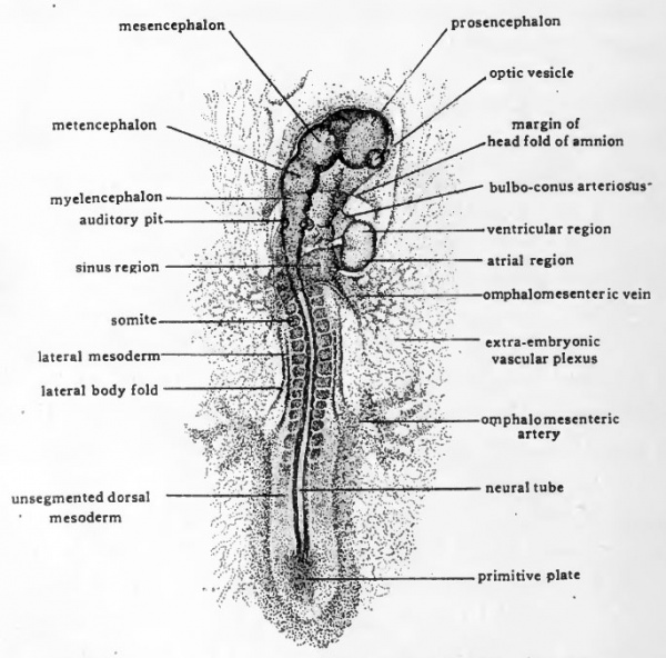
Torsion begins in the cephalic region of the embryo and progresses caudad. The first indications of torsion appear almost as soon as the cranial flexure begins and the two processes then progress synchronously. , In the chick, torsion is normally carried out toward a definite side. The cephalic region of the embryo is twisted in such a manner that the left side comes to lie next to the yolk and the right side away from the yolk. The progress of torsion caudad is gradual and the posterior part of the embryo remains prone on the yolk for a considerable time after torsion has been completed in the head region. Figure 22 shows the head of an embryo of about 38 hours in which the cranial flexure and torsion are just becoming evident. In chicks of about 43 hours (Fig. 29) the further progress of both flexion and torsion is well marked.
Fig. 29. Dorsal view ( X 14) of entire chick embryo having 19 pairs of somites (about 43 hours incubation). Due to torsion the cephalic region appears in dextro-dorsal view.
The processes of flexion and torsion thus initiated continue until the original orientation of the chick on the yolk is completely changed. As the body of the embryo becomes turned on its side the yolk no longer impedes the progress of flexion. Following the accomplishment of torsion in the cephalic region, the cranial flexure becomes rapidly greater until the head is practically doubled on itself (Fig. 34). As development proceeds, torsion progresses caudad involving more and more of the body of the embryo. Finally the entire embryo comes to lie with its left side on the yolk. Concomitant with the progress of torsion, flexion also appears farther caudally, affecting in turn the cervical, dorsal, and caudal regions. The series of flexions which accompany torsion bend the head and tail of the embryo ventrally so that its spinal axis becomes C-shaped (Fig. 40). The flexions which bend the embryo on itself so the head and tail lie close together are characteristic of all amniote embryos. The torsion which in the chick accompanies flexion is correlated with the fact that it develops on the surface of a large yolk.
The Completion of the Vitelline Circulatory Channels
In chicks of 33 to 36 hours the omphalomesenteric veins have been established as postero-lateral extensions of the same endocardial tubes which are involved in the formation of the heart. As the omphalomesenteric veins are extending laterad, the vessels developing in the vitelline plexus are extending and converging toward the embryo. Eventually the vitelline vessels attain communication with the heart by becoming confluent with the omphalomesenteric veins. This establishes the afferent channels of the vitelline circulation.
The vessels destined to carry blood from the embryo to the vitelline plexus develop in embryos of about 40 hours (Fig. 29). Like the afferent vitelline channels, the efferent channels have a dual origin. The proximal portions of the efferent channels arise within the embryo as branches of the dorsal aortae, and extend peripherally. The distal portions of the channels arise in the extra-embryonic vascular area and extend toward the embryo. The efferent vitelline vessels are estabhshed when these two sets of channels become confluent. In its early stages the connection is through a network of small channels rather than definite vessels, the aortae breaking up posteriorly into small channels some of which communicate laterally with the extra-embryonic plexus. Later some of these channels become confluent, others disappear, and gradually definite main vessels, the omphalomesenteric arteries, are established. For some time after their formation, the omphalomesenteric arteries are likely to retain traces of their origin from a plexus of small channels and arise from the aorta by several roots (Fig. 35).
The Beginning of the Circulation of Blood
At about 44 hours of incubation, coincident with the completion of the vitelline vessels, the heart begins regular contraction, and the blood which has been formed in the extra-embryonic vascular area is for the first time pumped through the vessels of the embryo. In tracing the course of either the embryonic or the vitelline circulation the heart is the logical starting point. From the heart the blood of the extra-embryonic vitelline circulation passes through the ventral aortae, along the dorsal aortae, and out through the omphalomesenteric arteries to the plexus of vessels on the yolk.
In the small vessels which ramify in the membranes enveloping the yolk the blood absorbs food material. In young embryos, before the allantoic circulation has appeared, the vitelline circulation is involved also in the oxygenation of the blood. The great surface exposure presented by the multitude of small vessels on the yolk makes it possible for the blood to take up oxygen which penetrates the porous shell and the albumen.
After acquiring food material and oxygen the blood is collected by the sinus terminalis and the vitelUne veins. The vitelline veins converge toward the embryo from all parts of the vascular area and empty into the omphalomesenteric veins which return the blood to the heart (Fig. 48).
The blood of the intra-embryoiiic circulation, leaving the heart enters the ventral aortae, thence passes into the dorsal aortae, and is distributed through branches from the dorsal aortae to the body of the embryo. It is returned from the cephalic part of the body by the anterior cardinals, and from the caudal part of the body by the posterior cardinals. The anterior and posterior cardinals discharge together through the ducts of Cuvier into the sinus region of the heart (Fig. 24) .
In the heart, the blood of the extra-embryonic circulation and of the intra-embryonic circulaSon is mixed. The mixed blood in the heart is not as rich in oxygen and food material as that which comes to the heart from the vitelline circulation nor as low in food and oxygen content as that returned to the heart from the intra-embryonic circulation where these materials are drawn upon by the growing tissues of the embryo. Nevertheless it carries a sufficient proportion of food and oxygen so that as it is distributed to the body of the embryo it serves to supply the growing tissues.
The Early Embryology of the Chick: Introduction | Gametes and Fertilization | Segmentation | Entoderm | Primitive Streak and Mesoderm | Primitive Streak to Somites | 24 Hours | 24 to 33 Hours | 33 to 39 Hours | 40 to 50 Hours | Extra-embryonic Membranes | 50 to 55 Hours | Day 3 to 4 | References | Figures | Site links: Embryology History | Chicken Development
| Historic Disclaimer - information about historic embryology pages |
|---|
| Pages where the terms "Historic" (textbooks, papers, people, recommendations) appear on this site, and sections within pages where this disclaimer appears, indicate that the content and scientific understanding are specific to the time of publication. This means that while some scientific descriptions are still accurate, the terminology and interpretation of the developmental mechanisms reflect the understanding at the time of original publication and those of the preceding periods, these terms, interpretations and recommendations may not reflect our current scientific understanding. (More? Embryology History | Historic Embryology Papers) |
Glossary Links
- Glossary: A | B | C | D | E | F | G | H | I | J | K | L | M | N | O | P | Q | R | S | T | U | V | W | X | Y | Z | Numbers | Symbols | Term Link
Cite this page: Hill, M.A. (2024, April 30) Embryology Book - The Early Embryology of the Chick 10. Retrieved from https://embryology.med.unsw.edu.au/embryology/index.php/Book_-_The_Early_Embryology_of_the_Chick_10
- © Dr Mark Hill 2024, UNSW Embryology ISBN: 978 0 7334 2609 4 - UNSW CRICOS Provider Code No. 00098G


