Book - Text-Book of Embryology 14
| Embryology - 27 Apr 2024 |
|---|
| Google Translate - select your language from the list shown below (this will open a new external page) |
|
العربية | català | 中文 | 中國傳統的 | français | Deutsche | עִברִית | हिंदी | bahasa Indonesia | italiano | 日本語 | 한국어 | မြန်မာ | Pilipino | Polskie | português | ਪੰਜਾਬੀ ਦੇ | Română | русский | Español | Swahili | Svensk | ไทย | Türkçe | اردو | ייִדיש | Tiếng Việt These external translations are automated and may not be accurate. (More? About Translations) |
Bailey FR. and Miller AM. Text-Book of Embryology (1921) New York: William Wood and Co.
- Contents: Germ cells | Maturation | Fertilization | Amphioxus | Frog | Chick | Mammalian | External body form | Connective tissues and skeletal | Vascular | Muscular | Alimentary tube and organs | Respiratory | Coelom, Diaphragm and Mesenteries | Urogenital | Integumentary | Nervous System | Special Sense | Foetal Membranes | Teratogenesis | Figures
| Historic Disclaimer - information about historic embryology pages |
|---|
| Pages where the terms "Historic" (textbooks, papers, people, recommendations) appear on this site, and sections within pages where this disclaimer appears, indicate that the content and scientific understanding are specific to the time of publication. This means that while some scientific descriptions are still accurate, the terminology and interpretation of the developmental mechanisms reflect the understanding at the time of original publication and those of the preceding periods, these terms, interpretations and recommendations may not reflect our current scientific understanding. (More? Embryology History | Historic Embryology Papers) |
The development of the coelom, the pericardium, pleuroperitoneum, diaphragm and mesenteries
In the Chapter on the development of the germ layers, it is stated that the peripheral part of the mesoderm splits into two layers, an outer or parietal, and an inner or visceral (Fig. 72; see also p. 96). The parietal layer of mesoderm and the ectoderm constitute the somatopleure. The visceral layer and the entoderm constitute the splanchnopleure (Fig. 72). The cleft or cavity that appears between the parietal and visceral layers is the coelom or body cavity and is lined with a layer of flattened mesodermal cells known as the mesothelium. It will be remembered that in the earlier stages of development a portion of the embryonic disk becomes constricted off from the yolk sac to form the simple cylindrical body (p. 107) . Along each side of the axial portion of the germ disk, and also at its cephalic and caudal ends, the germ layers bend ventrally and then medially until they meet and fuse in the midventral line (p. 109) . In this way a part of the somatopleure forms the lateral and ventral portions of the body wall (Fig. 103). At the same time the axial portion of the entoderm is bent into a tube which is closed at both ends the primitive gut and is then pinched off from the rest of the entoderm except at one point, where the cavity of the gut remains in communication with the cavity of the yolk sac. The splanchnic mesoderm adjacent to the entoderm on each side comes in contact and fuses with the corresponding portion from the opposite side, thus forming a sheet of tissue which encloses the primitive gut and also forms a partition between the two parts of the coelom. This sheet of tissue is the common mesentery and is attached to the dorsal and ventral body walls along the medial line. The portion between the gut and the dorsal body wall is the dorsal mesentery, the portion between the gut and the ventral body wall is the ventral mesentery. Thus the gut is suspended in the common mesentery (Figs. 197 and 282).
When portions of the somatopleure and splanchnopleure are bent ventrally the coelom between the portions is naturally carried with them. This part of the coelom thus becomes enclosed within the cylindrical body and constitutes the intraembryonic or simply the embryonic codom (body cavity proper). The part of the ccelom which, while the germ layers were still flat, was situated more peripherally constitutes the extraembryonic coelom or eococcdom (extraembryonic body cavity). From the nature of the bending process, the embryonic coelom is divided into bilaterally symmetrical parts by the common mesentery (Fig. 197). These two simple cavities are the forerunners of all the serous cavities of the body. The various partitions between the serous cavities, the walls of the cavities and the mesenteries proper are all derived from the somatic and splanchnic mesoderm with its covering of mesothelium.
While the foregoing would represent a typical case of early ccelom and mesentery formation, there are certain modifications and peculiarities in the higher Mammals and in man. In all cases the splitting of the mesoderm to form the coelom proceeds from the periphery of the germ disk toward the axial portion (p. 80) . In the human embryo the bending ventrally and fusing of the germ layers to form the cylindrical body begins in the anterior region of the disk and is accomplished there before the splitting of the mesoderm is completed. The peripheral splitting has resulted in the formation of the exoccelom, but at the time when the ventral fusion of the germ layers takes place, the splitting has not extended axially to a sufficient degree to form the intraembryonic crelom. The latter, which appears later in this region, never communicates laterally, therefore, with the exocoelom. Caudal to this region the coelom is formed as in the typical case. The more anterior part of the coelom on each side is thus primarily a pocket-like cavity. It communicates with the rest of the coelom at about the level of the yolk stalk. In the region of the fore-gut, the future oesophagus, no distinct mesentery is formed, but the fore-gut remains broadly attached to the dorsal body wall. A ventral mesentery is lacking from a point just cranial to the yolk stalk to the caudal end of the gut. There are no coelomic cavities in the branchial arches, the ccelom extending only to the last branchial groove.
In very young human embryos the primitive segments contain small cavities. These cavities soon disappear, being filled with cells from the surrounding, parts of the segments. Whether they represent isolated portions of the coelom is not certain. In the lower Vertebrates, the cavities of the primitive segments regularly communicate with the ccelom, and in the sheep the cavities of the first formed segments are continuous with the ccelom. In the head there is no cavity analogous to the coelom in the body. In but one human embryo have any cavities in the head resembling those of the primitive segments been observed (see p. 2 70) .
The Pericardial Cavity, Pleural Cavities and Diaphragm
The pericardial and pleural cavities and diaphragm are so closely related in their development that they must be considered together. In the region just caudal to the visceral arches, where the two anlagen of the heart appear, the embryonic coelom becomes dilated at a very early stage to form the primitive pericardial cavity (parietal cavity of His). After the two anlagen of the heart unite to form a simple tubular structure (p. 196: also Fig. 156), the latter is suspended in the cavity by a mesentery which consists of a dorsal and a ventral part, a dorsal and a ventral mesocardium. By these the cavity is at first divided into two bilaterally symmetrically parts. The mesocardia soon disappear and leave the heart attached only to the large vascular trunks which suspend it in the single pericardial cavity. The early pericardial cavity is simply the cephalic end of the embryonic coelom and is therefore directly continuous with the rest of the coelom. As mentioned on p. 341 it does not, however, at any time communicate laterally with the extraembryonic coelom.
The communication between the pericardial cavity and the rest of the embryonic coelom is soon partly cut off by the development of a transverse fold - the septum transversum. This septum is formed in close relation with the omphalomesenteric veins. These vessels unite in the sinus venosus at the caudal end of the heart, whence they diverge in the splanchnic mesoderm.
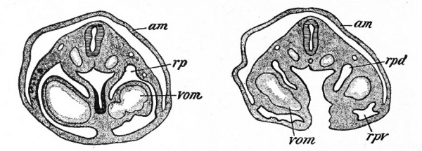
Fig. 291. Transverse sections of a rabbit embryo Showing how the omphalomesenteric veins (vom) push outward across the coelom and fuse with the lateral body wall, forming the ductus pleuro-pericardiacus (rp, rpd) ; am, amnion. Ravn.
They are thus embedded in the mesodermal layer of the splanchnopleure, and as the latter closes in from either side to form the gut, the vessels form ridge-like projections into the coelom. As the vessels increase in size, the ridges become so large that the splanchnic mesoderm is pushed outward against the parietal mesoderm and fuses with it (Fig. 291). Thus a partition is formed on each side, which is attached on the one hand to the mesentery and on the other hand to the ventral and lateral body walls, and which contains the omphalomesenteric veins. It is obvious that these partitions, forming the septum transversum, close the ventral part of the communication between the pericardial cavity and the rest of the coelom. The dorsal part of the communication remains open on each side of the mesentery as the ductus pleuro-pericardiacus (dorsal parietal recess of His) (Figs. 291 and 292).
As the heart develops it migrates caudally, and by corresponding migration the pericardial cavity draws the ventral edge of the septum transversum farther caudally, so that the cephalic surface of the latter faces ventrally and cranially.
In other words the septum comes to lie in an oblique cranio-caudal plane. The pericardial cavity therefore comes to lie ventral to the ductus pleuro-pericardiaci. The latter one on each side of the mesentery are two passages which communicate on the one hand with the pericardial cavity and on the other hand with the peritoneal cavity, while they themselves form the cavities into which the lungs grow the pleural cavities. (Compare Figs. 292, 293 and 294.)
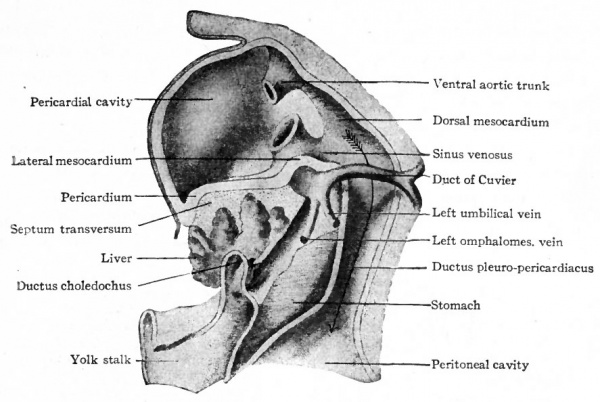
Fig. 292. From a model of the septum transversum, liver etc., of a human embryo of 3 mm. His, Kollmann.
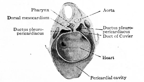
Fig. 293. View (in perspective) of the pericardial cavity and ductus pleuro-pericardiaci of a rabbit embryo of 9 days. Ravn.
The pleural cavities also become separated from the pericardial cavity, apparently through the agency of the ducts of Cuvier. The anterior and posterior cardinal veins on each side unite to form the duct of Cuvier which then extends from the body wall through the dorsal free edge of the septum transversum to join the sinus venosus (Fig. 292). This free edge is pushed farther and farther into the ductus pleuro-pericardiacus (Fig. 293) until it meets and fuses with the mesentery or posterior mediastinum. This process thus produces a septum between each pleural cavity and the pericardial cavity.
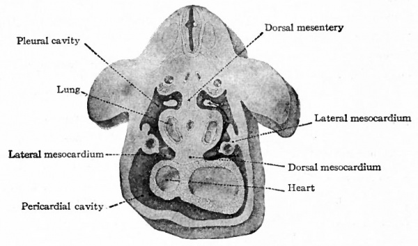
Fig. 294. View (in perspective) of the pericardial and pleural cavities of a human embryo of 7.5 mm. Kollmann.
- The arrow points through the opening which forms the communication between the pleural and peritoneal cavities, and which is eventually closed by the pleuro-peritoneal membrane.
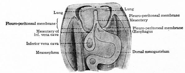
Fig. 295. Ventral view (in perspective) of parts of the lungs, pleural cavities, peritoneal cavity, and the pleuro-peritoneal membranes in a rat embryo. Ravn.
The septum transversum early acquires still more complicated relations from the fact that the liver grows into its caudal part (Fig. 292) . It may, for this reason, be divided into a caudal part in which the liver is situated and which furnishes the fibrous capsule (of Glisson) and the connective tissue of the liver, and a cephalic part which may be called the primary diaphragm. These two parts at first are not separate, the separation taking place secondarily. After the separation between the pericardial cavity and the pleural cavities, the latter for a time remain in open communication with the rest of the coelom or peritoneal cavity. The lungs, as they develop, grow into the pleural cavities (Fig. 294) until their tips finally touch the cephalic surface of the liver. At this point folds grow from the lateral and dorsal body walls (Fig. 295) and unite ventrally with the primary diaphragm and medially with the mesentery. These folds the pleuroperitoneal membranes separate the pleural cavities from the peritoneal cavity and complete the diaphragm. Thus the diaphragm, from the standpoint of development, consists of two parts : a ventral part which is the cephalic portion of the original septum transversum, and a dorsal part which develops later from the body wall and is the closing membrane between the peritoneal and pleural cavities. The musculature of the diaphragm is considered in the chapter on the muscular system (p. 269).
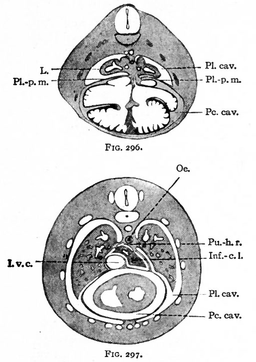
Fig. 296. Transverse section through the thoracic region of a rabbit embryo of 15 days. Hochstetter.
Fig. 297. Transverse section through the thoracic region of a cat embryo of 25 mm. Hochstetter.
- I.v.c.. Inferior vena cava; Inf.-c. 1., infracardiac lobe of lung; L., lung; Oe.. oesophagus; PC. cav. t pericardial cavity; PI. cav., pleural cavity; Pl.-p. m., pleuro- pericardial membrane; Pu.-h. r., pulmo-hepatic recess.
While the foregoing structures are being formed, decided changes take place in their positions and relations. At first the heart lies far forward in the cervical region near the visceral arches. Later it migrates caudally and the pericardial cavity comes to occupy much of the ventral part of the thorax, the pericardium having extensive attachments to the ventral body wall and to the cephalic surface of the primary diaphragm (Fig. 292). The diaphragm is much farther forward than in the adult and is broadly attached to the cephalic surface of the liver. The principal changes which bring about the adult conditions are the growth of the lungs, the separation of the diaphragm from the liver, and the caudal migration of the diaphragm itself. With the development of the lungs, the pleura! cavities necessarily enlarge and push their way ventrally. In so doing they split the pericardium away from the lateral body walls and likewise from the diaphragm (compare Figs. 296 and 297). Thus the pericardial cavity comes to be confined more and more closely to the medial ventral position. The separation of the liver from the primary diaphragm is caused by changes in the peritoneum which at first covers the caudal, lateral and ventral surfaces of the liver. The cephalic surface of the liver, as stated above, is covered by the primary diaphragm itself. The peritoneum is reflected from the surface of the liver on to the diaphragm, and at the line of reflection a groove appears on each side, extending from the midventral line around as far as the attachment of the liver to the diaphragm.
The grooves gradually grow deeper, the peritoneum rum^embry^T^K Pushing its way, as a flat sac, between the two stages. structures, until the separation is almost complete.
This is a remnant of the ventral mesentery and forms the ligamentum suspensorium (falciforme) hepatis. In its free caudal edge: is embedded the ligamentum teres hepatis which is closely related to the! umbilical vein (see p. 230). The diaphragm itself, during its development,, migrates from a position in the cervical region, where the septum transversuin first appears, to its final position opposite the last thoracic vertebrae. During: the migration the plane of direction also changes several times, as may be seen in Fig. 298.
Fig. 298. Diagram showing the position of the diaphragm in human embryos of different stages. Mall. The positions are those shown in embryos of Mall's collection (except KO, which is a 10.2 mm embryo of the His collection); XII being an embryo 2.1 mm; XVIII of 7mm; XIX of 5 mm; II of 7mm; IX of 17mm; XLIII of 15mm; VI of 24mm. The numerals on the right indicate segments.
Pericardium, Pleuroperitoneum, Diaphragm and Mesenteries
The Pericardium and Pleura. Since the pericardial cavity represents a portion of the original ccelom, the lining of the cavity must be a derivative of either the parietal or the visceral layer of mesoderm or of both. The common mesentery in which the heart develops is derived from the visceral layer. Consequently the epicardium is a derivative of the visceral mesoderm (Fig. 165). The pericardium is derived from three regions of mesoderm. The greater part is derived from the parietal mesoderm, since the body wall which is composed of parietal mesoderm is also primarily the wall of the pericardial cavity. A small dorsal portion is probably derived from the mesoderm at the root of the dorsal mesocardium (Fig. 165). The septum transversum primarily forms the caudal wall of the pericardial cavity, and, since the septum is a derivative of the visceral layer, the caudal wall is derived from this layer. The three portions are, of course, Continuous.
The lungs first appear in the common mesentery as an evagination from the primitive gut (Fig. 282, p. 330;. As they develop further they grow into the pleural cavities, pushing a part of the mesentery before them. This part of the mesentery thus invests the lungs and forms the visceral layer of the pleura which is therefore a derivative of the visceral mesoderm. The parietal layer of the pleura is a derivative of the parietal mesoderm, since the wall of the pleural cavity is primarily the body wall.
The lining of all these cavities is at first composed of mesothelium and mesenchyme. The latter is transformed into the delicate connective tissue of the serous membranes, and the mesothelium becomes the mesothelium of the membranes.
The Omentum and Mesentery
From the septum transversum (or diaphragm) to the anus the gut is suspended in the ccelom (or abdominal cavity) by means of the dorsal mesentery. This is attached to the dorsal body wall along the medial line and lies in the medial sagittal plane (Fig. 263 ; compare with Fig. 197). On the ventral side of the gut a mesentery is lacking from the anus to a point just cranial to the yolk ; stalk (p. 341). There is, however, a small ventral mesentery extending a short j distance caudally from the septum transversum. On account of its relation to I the stomach this is known as the ventral mesogastrium (Fig. 263). These two | sheets of tissue, the dorsal and ventral mesenteries, are destined to give rise to ; the omenta and mesenteries of the adult. Owing to the enormous elongation of the gut and its extensive coiling in the abdominal cavity, the primary mesenteries are twisted and thrown into many folds which enclose certain pockets or i bursae. Furthermore, certain parts of the gut which are originally free and movable become attached to other parts and to the body walls through fusions of certain parts of the mesentery with one another and with the body walls.
The Greater Omentum and Omental Bursa
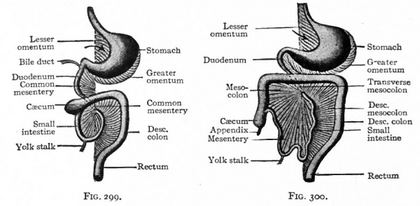
A small part of the gut caudal to the diaphragm is destined to become the stomach, and the portion of the mesentery which attaches it to the dorsal body wall is known as the dorsal mesogastrium (Fig. 263). The latter is inserted along the greater curvature of the stomach and lies in the medial sagittal plane so long as the stomach lies in this plane. When the stomach turns so that its long axis lies in a transverse direction and its greater curvature is directed caudally (p. 305), the dorsal mesogastrium changes its position accordingly. From its attachment along the dorsal body. wall it bends to the left and then ventrally to its attachment along the greater curvature of the stomach. Thus a sort of sac is formed dorsal to the stomach (Figs. 299 and 300). This sac is really a part of the abdominal or peritoneal cavity and opens toward the right side. The ventral wall is formed by the stomach, the dorsal and caudal walls by the mesogastrium. The cavity of the sac is the omental bursa (bursa omentalis) ; the mesogastrium forms the greater omentum (omentum majus) . The opening from the bursa into the general peritoneal cavity is the epiploic foramen (foramen of Winslow). (Fig. 276.)
Fig. 299. Diagram of the gastrointestinal tract and its mesenteries at an early stage. Ventral view. Hertwig.
Fig. 300. Same at a later stage. Hertwig. The arrow points into the bursa omentalis.
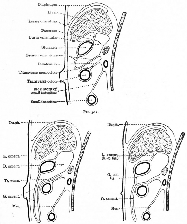
From the third month on, the greater omentum becomes larger and gradually extends toward the ventral abdominal wall, over the transverse colon, and then caudally between the body wall and the small intestine (Figs. 301 and 302). The portion between the body wall and intestine encloses merely a flat cavity continuous with the larger cavity dorsal to the stomach. From the fourth month on, the omentum fuses with certain other structures and becomes less free. The dorsal lamella fuses with the dorsal body wall on the left side and with the transverse mesocolon and transverse colon (Fig. 303). During the first or second year after birth the two lamellae fuse with each other caudal to the transverse colon to form the greater omentum of adult anatomy.
Figs. 301, 302 and 303. Diagrams showing stages in the development of the bursa omentalis, the greater omentum, and the fusion of the latter with the transverse mesocolon. Diagrams represent sagittal sections. For explanation of lettering in Figs. 302 and 303 see Fig. 301.
The Lesser Omentum
It has already been noted that the liver grows into the caudal portion of the septum transversum (p. 344). Since the ventral mesentery in the abdominal region, or the ventral mesogastrium, is primarily directly continuous with the septum transversum, it is later attached to the liver. In other words it passes between the liver and the lesser curvature of the stomach and also extends along the duodenal portion of the gut for a short distance (Fig. 263). As the stomach turns to the left the ventral mesentery is also drawn toward the left and comes to lie almost at right angles to the sagittal plane of the body, forming the lesser omentum (omentum minus) or the hepatogastric and hepatoduodenal ligaments of the adult (Figs. 303 and 304).
The Mesenteries
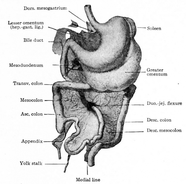
So long as the intestine is a straight tube, the dorsal mesentery lies in the medial sagittal plane, its dorsal attachment being practically of the same length as its ventral (intestinal) attachment. As development proceeds, the intestine elongates much more rapidly than the abdominal walls, and the intestinal attachment of the mesentery elongates accordingly. When the portion of the intestine to which the yolk stalk is attached grows out into the proximal end of the umbilical cord (p. 307) , the corresponding portion of the mesentery is drawn out with it (Fig. 263). When the intestine returns to the abdominal cavity and forms the primary loop, with the caecum to the right side (p. 308), its mesenteric attachment is carried out of the medial sagittal plane. This results in a funnel-shaped twisting of the mesentery (Figs. 299 and 300). The portion of the mesentery which forms the funnel is destined to become the mesentery of the jejunum, ileum, and ascending and transverse colon, and is attached to the dorsal body wall at the apex of the funnel (Figs. 299 and 300, 304). This condition is reached about the middle of the fourth month.
Up to this time the mesentery and intestine are freely movable, that is, they have formed no secondary attachments. From this time on, as the intestine continues to elongate and forms loops and coils, the mesentery is thrown into folds, and certain parts of it fuse with other parts and with the body wall. Thus certain parts of the intestine become less free or less movable within the abdominal cavity.
The duodenum changes from the original longitudinal position to a more nearly transverse position and, with its mesentery the mesoduodenum fuses with the dorsal body wall, thus becoming firmly fixed. Since the mesoduodenum fuses with the body wall, the duodenum has no mesentery in the adult. The pancreas, which is primarily enclosed within the mesoduodenum, also becomes firmly attached to the dorsal body wall (compare Figs. 301 and 302).
The mesentery of the transverse colon, or the transverse mesocolon, which lies across the body ventral to the duodenum (Figs. 300 and 304), fuses with the ventral surface of the latter and with the peritoneum of the dorsal body wall. In this way the dorsal attachment of the transverse mesocolon is changed from its original sagittal direction to a transverse direction (Figs. 302 and 303). The mesocolon itself forms a transverse partition which divides the peritoneal cavity into two parts, an upper (or cranial) which contains the stomach and liver, and a lower (or caudal) which contains the rest of the digestive tube except the duodenum. The mesentery of the duodenum and pancreas changes from a serous membrane into subserous connective tissue, and these two organs assume the retroperitoneal position characteristic of the adult (Fig. 301).
The mesentery of the descending colon, or the descending mesocolon, lies in the left side of the abdominal cavity, in contact with the peritoneum of the body wall (see Fig. 304). It usually fuses with the peritoneum, and the descending colon thus becomes fixed. After the ascending colon is formed, the ascending mesocolon usually fuses with the peritoneum on the right side (see Fig. 304). In a large percentage (possibly 25 per cent.) of individuals, the fusion between the peritoneum and the ascending and descending mesocolon is incomplete or wanting.
Fig. 304. Gastrointestinal tract and mesenteries in a human embryo. The arrow points into the bursa omentalis. Kollmann.
The sigmoid mesocolon bends to the left to reach the sigmoid colon, but forms no secondary attachments. It is continuous with the mesorectum which maintains its original sagittal position. A sheet of tissue the mesoappendix continuous with and resembling the mesentery, is attached to the caecum and vermiform appendix (Fig. 304). It probably represents a drawn out portion of the original common mesentery, since the caecum and appendix together are formed as an evagination from the primitive gut.
Normally the mesentery of the small intestine forms no secondary attachments, but is thrown into a number of folds which correspond to the coils of the intestine.
The secondary attachments of the greater omentum and the fusion of the two lamellae have been described earlier in this chapter (p. 348) . The mesenteries of the urogenital organs are considered in connection with the development of those organs (Chapter XV).
The Peritoneum. The thin layer of tissue composed of delicate fibrous connective tissue and mesothelium which lines the abdominal cavity and is reflected over the various omenta, mesenteries and visceral organs, is derived wholly from the mesoderm. The lining of the ccelom is composed of mesothelium and mesenchyme. The latter gives rise to the connective tissue of the serous membranes, and the mesothelial layer becomes the mesothelium of these membranes.
Anomalies
The Pericardium
Anomalous conditions of the pericardium are usually, although not necessarily, associated with anomalies of the heart. They may also be associated with defects in the diaphragm. Displacement of the heart (ectopia cordis) is accompanied by displacement of the pericardium. The heart sometimes protrudes through the thoracic wall, and, as a rule, in such cases is covered by the protruding pericardium. In extensive cleft of the thoracic wall (thoracoschisis, Chap. XX) the pericardium may be ruptured.
The Diaphragm
The most common malformation of the diaphragm is a defect in its dorsal part, occurring much more frequently on the left than on the right side. The defect may affect but a small portion or may be extensive, the peritoneum being directly continuous with the parietal layer of the pleura. Such defects are due to the imperfect development of the pleuro-peritoneal membrane which normally grows from the dorso-lateral part of the body wall and fuses with the edge of the primary diaphragm, thus separating the pleural and and peritoneal cavities (p. 345) . The most conspicuous result of defects in the dorsal part of the diaphragm is diaphragmatic hernia, in which parts of the stomach, liver, spleen and intestine project into the pleural cavity, either free or enclosed in a peritoneal sac. Defects in the ventral part of the diaphragm, due to imperfect development of portions of the septum transversum, are not common.
The Mesenteries and Omenta
Extensive malformations of the mesenteries apparently do not occur without extensive malformations of the digestive tract. One of the most striking anomalous conditions is a retained embryonic simplicity of the mesentery, concurrent with corresponding simplicity in the loops and coils of the intestine. In this anomaly the intestine has failed to arrive at its usual complicated condition and the mesentery has not undergone the usual processes of folding and fusion (p. 350 et seq.). Minor variations in the mesenteries and omenta are probably due largely to imperfect fusion of certain parts with one another and with the body wall. It is not uncommon to find the ascending or descending colon, or both, more or less free and movable. This condition is due to imperfect fusion of the mesocolon with the body wall (p. 351). If the greater omentum is wholly or partially divided into sheets of tissue, the two primary lamellae have failed to fuse completely (p. 349). This divided condition is normal in many Mammals.
- Next: Urogenital
References for Further Study
ib BRACKET, A.: Recherches sur le developpement du diaphragme et du foie. Jour, de
VAnat. et de la Physiol., T. XXXII, 1895.
BROMAN, J.: Die Entwickelungsgeschichte der Bursa omentalis und ahnlicher Recess* bildungen bei den Wirbeltieren. Wiesbaden, 1904.
BROMAN, I.: Ueber die Entwickelung und Bedeutung der Mesenterien und der Korperhohlen bei den Wirbeltieren. Ergebnisse der Anat. u. Entwick., Bd. XV, 1906.
BROSSIKE, G.: Ueber intraabdominale (retroperitoneale) Hernien und Bauchfelltaschen, nebst einer Darstellung der Entwickelung peritonealer Formationen. Berlin, 1891.
HERTWIG, O. : Lehrbuch der Entwickelungsgeschichte des Menschen und der Wirbeltiere. Jena, 1906.
KEIBEL, F., and MALL, F. P.: Manual of Human Embryology, Vol. I, 1910.
KLAATSCH: Zur Morphologic der Mesenterialbildungen am Darmkanal der Wirbeltiere. Morph. Jahrbuch, Bd. XVIII, 1892.
KOLLMANN, J.: Lehrbuch der Entwickelungsgeschichte des Menschen. Jena, 1898.
KOLLMANN, J.: Handatlas der Entwickelungsgeschichte des Menschen. Bd. II, 1907.
MALL, F. P.: Development of the Human Ccelom. Jour, of Morphol., Vol. XII, 1897.
MALL, F. P.: On the Development of the Human Diaphragm. Johns Hopkins Hospital Bulletin, Vol. XII, 1901.
PIERSOL. G. A.: Teratology. In Wood's Reference Handbook of the Medical Sciences. 1904.
RAVN, E.: Ueber die Bildung der Scheidewand zwischen Brust- und Bauchhohle in Saugetierembryonen. Arch. f. Anat. u. Physiol., Anat. Abth., 1889.
STRAHL and CARIUS: Beitrage zur Entwickelungsgeschichte des Herzens und der Korperhohlen. Arch. /. Anat. u. Physiol., Anat. Abth., 1889.
SWAEN, A.: Recherches sur le developpement du foie, du tube digestif, de 1'arrierecavite du peritoine et du mesentere. Premiere partie, Lapin. Jour, de VAnat. et de la Physiol., T. XXXIII, 1896. Seconde partie. Embryons humains. T. XXXIII, 1897.
TOLDT, C.: Bau und Wachstumsveranderung der Gekrose des menschlichen Darmkanals. Denkschr. der kais. Akad. Wissensch. Wien. Math.-Naturwissen. Classe, Bd XLI, 1879.
| Historic Disclaimer - information about historic embryology pages |
|---|
| Pages where the terms "Historic" (textbooks, papers, people, recommendations) appear on this site, and sections within pages where this disclaimer appears, indicate that the content and scientific understanding are specific to the time of publication. This means that while some scientific descriptions are still accurate, the terminology and interpretation of the developmental mechanisms reflect the understanding at the time of original publication and those of the preceding periods, these terms, interpretations and recommendations may not reflect our current scientific understanding. (More? Embryology History | Historic Embryology Papers) |
Text-Book of Embryology: Germ cells | Maturation | Fertilization | Amphioxus | Frog | Chick | Mammalian | External body form | Connective tissues and skeletal | Vascular | Muscular | Alimentary tube and organs | Respiratory | Coelom, Diaphragm and Mesenteries | Urogenital | Integumentary | Nervous System | Special Sense | Foetal Membranes | Teratogenesis | Figures
Glossary Links
- Glossary: A | B | C | D | E | F | G | H | I | J | K | L | M | N | O | P | Q | R | S | T | U | V | W | X | Y | Z | Numbers | Symbols | Term Link
Cite this page: Hill, M.A. (2024, April 27) Embryology Book - Text-Book of Embryology 14. Retrieved from https://embryology.med.unsw.edu.au/embryology/index.php/Book_-_Text-Book_of_Embryology_14
- © Dr Mark Hill 2024, UNSW Embryology ISBN: 978 0 7334 2609 4 - UNSW CRICOS Provider Code No. 00098G

