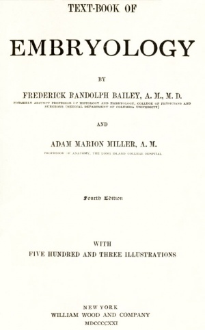
Book - Text-Book of Embryology (1921)
| Embryology - 27 Apr 2024 |
|---|
| Google Translate - select your language from the list shown below (this will open a new external page) |
|
العربية | català | 中文 | 中國傳統的 | français | Deutsche | עִברִית | हिंदी | bahasa Indonesia | italiano | 日本語 | 한국어 | မြန်မာ | Pilipino | Polskie | português | ਪੰਜਾਬੀ ਦੇ | Română | русский | Español | Swahili | Svensk | ไทย | Türkçe | اردو | ייִדיש | Tiếng Việt These external translations are automated and may not be accurate. (More? About Translations) |
Bailey FR. and Miller AM. Text-Book of Embryology (1921) New York: William Wood and Co.
- Contents: Germ cells | Maturation | Fertilization | Amphioxus | Frog | Chick | Mammalian | External body form | Connective tissues and skeletal | Vascular | Muscular | Alimentary tube and organs | Respiratory | Coelom, Diaphragm and Mesenteries | Urogenital | Integumentary | Nervous System | Special Sense | Foetal Membranes | Teratogenesis | Figures
| Historic Disclaimer - information about historic embryology pages |
|---|
| Pages where the terms "Historic" (textbooks, papers, people, recommendations) appear on this site, and sections within pages where this disclaimer appears, indicate that the content and scientific understanding are specific to the time of publication. This means that while some scientific descriptions are still accurate, the terminology and interpretation of the developmental mechanisms reflect the understanding at the time of original publication and those of the preceding periods, these terms, interpretations and recommendations may not reflect our current scientific understanding. (More? Embryology History | Historic Embryology Papers) |
Preface to the Fourth Edition
In the present edition the plan of the book has been modified in certain respects. The chapter on the cell has been omitted because in the opinion of the authors the previous training of the student who commences the study, of the embryology of vertebrates has been sufficient to bring to his attention the salient features of cell organization. In former editions the early processes of development, viz: cleavage, gastrulation, and mesoderm formation, were treated as topics in separate chapters. The present plan comprises the treatment of the early stages in succession in a given animal form; individual chapters are devoted to Amphioxus, the frog, the chick, and the mammal. This change has been made because it is our opinion gained from experience in teaching that the student acquires a better understanding of the development of the germ layers by following the processes as a continuous series in a given animal. A number of old illustrations have been replaced by new figures the sources of which have been duly credited.
Apart from the insertion of the chapter on foetal membranes the second part of the book, comprising organogeny, has been revised only in so far as the results of recent investigation have modified the ideas expressed in the previous edition.
We wish to express our appreciation of the helpful criticisms of our colleagues and other friends.
The Authors. July, 1921.
Preface to the First Edition
The Text-book, as originally planned, is an outgrowth of the course in Embryology given at the Medical Department of Columbia University. It was intended primarily to present to the student of medicine the most important facts of development, at the same time emphasizing those features which bear directly upon other branches of medicine. As the work took form, it seemed best to broaden its scope and make it of greater value to the general student of embryology and allied sciences. With the opinion that illustrations convey a much clearer conception of structural features than verbal description alone, the writers have made free use of figures.
The plan of adding brief "Practical Suggestions" at the end of each chapter has been so thoroughly satisfactory in the Text-book of Histology, especially in connection with laboratory work, that it has been adopted here. These "suggestions" are not intended to be complete descriptions of embryological technic, but are for the purpose of furnishing the laboratory worker with certain of the more essential practical hints for studying the structures described in the chapter. To avoid frequent repetition, some of the best methods of procuring, handling, and preparing embryological material, and some of the more important formulae are given in the Appendix, which is intended to be used mainly for the carrying out of the "Practical Suggestions."
The development of the Germ Layers has been treated rather elaborately from a comparative standpoint, because this has been found the most satisfactory method of teaching the subject.
In the chapter on the Nervous System the aim has been to give a general conception of the subject, which, if once mastered by the student, will give him an insight into the structure and significance of the nervous system that will bring this difficult subject more fully within his grasp.
In Part II (Organogenesis), at the end of each chapter there is given a brief description of certain developmental anomalies which may occur in connection with the organs described in the chapter. In Chapter XIX (Teratogenesis) the nature and origin of the more complex anomalies and monsters are discussed, and also the causes underlying the origin of malformations.
The writers wish to thank Dr. Oliver S. Strong for his painstaking work on the chapter on the Nervous System. Dr. Strong in turn wishes to acknowledge his indebtedness to Dr. Adolf Meyer for important ideas underlying the treatment of his subject, and also for many valuable details. He expresses his thanks also to Professors C. J. Herrick, H. von W. Schulte and G. L. Streeter for helpful criticisms and suggestions. The writers would also express their thanks to Dr. H. McE. Knower for helpful criticisms on Part I and the chapter on Teratogenesis; to Dr. Edward Learning for making the photographs reproduced in the text; to the American Journal of Anatomy for the loan of plates; and to Messrs. William Wood & Company for their uniform courtesy and kindness.
Frederick Randolph Bailey. (1871 - 1923)
Adam Marion Miller.
April 1, 1909.
Contents
- The germ cells
- Maturation
- Fertilization
- Early development of amphioxus
- Early development of the frog
- Early development of the chick
- Early mammalian development
- Development of the external form of the body
- The development of connective tissues and the skeletal system
- The development of the vascular system
- The development of the muscular system
- The development of the alimentary tube and appended organs
- The development of the respiratory system
- The development of the coelom, the pericardium, pleuroperitoneum, diaphragm and mesenteries
- The development of the urogenital system
- The development of the integumentary system
- The nervous system
- The organs of special sense
- Foetal membranes
- Teratogenesis
Introduction
While Embryology as a science is of comparatively recent date, recorded observations upon the development of the foetus date back as far as 1600 when Fabricius ab Aquapendente published an article entitled "De Formato Foetu.' ; Four years later the same author added some further observations under the title, " De Formatione Foetus." Harvey (1651), using a simple lens, studied and described the chick embryo of two days' incubation. Harvey's idea was that the ovum consisted of fluid in which the embryo appeared by spontaneous generation. Regnier de Graaf (1677) described the ovarian follicle (Graafian follicle), and in the same year was announced the discovery by Von Loewenhoek of the spermatozoon. These and other embryologists of this period held what is now known as the prejormation theory. According to this theory, the adult form exists in miniature in the egg or germ, development being merely an enlarging and unfolding of preformed parts. With the discovery of the spermatozoon the " pref ormationists " were divided into two schools, one holding that the ovum was the container of the miniature individual (ovists), the other according this function to the spermatozoon (animalculists). According to the ovists, the ovum needed merely the stimulation of the spermatozoon to cause its contained individual to undergo development, whereas the animalculists looked upon the spermatozoon as the essential embryo-container, the ovum serving merely as a suitable food-supply or growing-place.
Nearly a hundred years of almost no further progress in embryological knowledge came to a close with the publication of Wolff's important article, "Theoria Generationis," in 1759. Wolff's theory was theory pure and simple, with very little basis on then known facts, but it was significant as being apparently the first clear statement of the doctrine of epigenesis. The two essential points in Wolff's theory were: (i) that the embryo was not preformed; that is, did not exist in miniature in the germ, but developed from a more or less unformed germ substance; (2) that union of male and female substances was necessary to initiate development. The details of Wolff's theory were wrong in that he looked upon the ovum as a structureless substance and upon the seminal fluid and not upon the spermatozoon as the male fecundative agent. Dollinger and his two pupils, von Baer and Pander, were the next to make important contributions to Embryology. Von Baer's publication in 1829 was of extreme significance in the development of embryological knowledge, for in it we have the first definite description of the primary germ layers as well as the first accurate differentiation between the Graafian follicle and the ovum. It will be remembered that the cell was not as yet recognized as the unit of organic structure. Only comparatively gross Embryology was thus possible. With the recognition of the cell as the basis of animal structure (Schleiden and Schwann, 1839) the entire field of histogenesis was opened to the embryologist; the ovum became known as a typical cell, while a little later (Kolliker, Reichert and others, about 1840) was established the function of the spermatozoon and the fact that it also was a modified cell structure. From this time we may consider the two fundamental facts of Histology and of Embryology, respectively, as firmly fixed beyond controversy; for Histology, the fact that the body consists wholly of cells and cell derivatives; for Embryology, the fact that all of these cells and cell derivatives develop from a single original cell the fertilized ovum.
The adult body being thus composed of an enormous number of cells, varying in structure and in function, forming the different tissues and organs, and these cells having all developed from the single fertilized germ cell, it is the province of Embryology to trace this development from the union of male and female germ cells to the cessation of developmental life.
While Embryology thus properly begins with the fertilized ovum, that is, with the first cell of the new individual, certain preliminary considerations are essential to the proper understanding of this cell and its future development. These are the structure of the ovum and of the spermatozoon and their development preparatory to union. Also, as it is with cells and cell activities that Embryology has largely to deal, it is necessary to consider the structure of the typical animal cell and the processes by which cells undergo division or proliferation.
While the subject of this work is distinctly human Embryology, it is neither possible nor advisable to confine our study wholly to human material. It is not possible, for the reason that material for the study of the earliest stages in the human embryo (first 12 days) is entirely wanting, while human embryos of under 20 days are extremely rare. Again, even later stages in human development are often best understood by comparison w r ith similar stages in lower forms. For practical study by the student, human material for all even of the later stages is rarely available, so that recourse must frequently be had to material from lower animals. Such study is, however, usually thoroughly satisfactory if the student has sufficient knowledge of comparative anatomy, and the deductions regarding human development, from the study of development in lower forms, are rarely in error.
- Next: Germ cells
| Historic Disclaimer - information about historic embryology pages |
|---|
| Pages where the terms "Historic" (textbooks, papers, people, recommendations) appear on this site, and sections within pages where this disclaimer appears, indicate that the content and scientific understanding are specific to the time of publication. This means that while some scientific descriptions are still accurate, the terminology and interpretation of the developmental mechanisms reflect the understanding at the time of original publication and those of the preceding periods, these terms, interpretations and recommendations may not reflect our current scientific understanding. (More? Embryology History | Historic Embryology Papers) |
Text-Book of Embryology: Germ cells | Maturation | Fertilization | Amphioxus | Frog | Chick | Mammalian | External body form | Connective tissues and skeletal | Vascular | Muscular | Alimentary tube and organs | Respiratory | Coelom, Diaphragm and Mesenteries | Urogenital | Integumentary | Nervous System | Special Sense | Foetal Membranes | Teratogenesis | Figures
Glossary Links
- Glossary: A | B | C | D | E | F | G | H | I | J | K | L | M | N | O | P | Q | R | S | T | U | V | W | X | Y | Z | Numbers | Symbols | Term Link
Cite this page: Hill, M.A. (2024, April 27) Embryology Book - Text-Book of Embryology (1921). Retrieved from https://embryology.med.unsw.edu.au/embryology/index.php/Book_-_Text-Book_of_Embryology_(1921)
- © Dr Mark Hill 2024, UNSW Embryology ISBN: 978 0 7334 2609 4 - UNSW CRICOS Provider Code No. 00098G

