Book - Manual of Human Embryology 6
Keibel F. VI. Summary of the development of the human embryo and the differentiation of its external form in Keibel F. and Mall FP. Manual of Human Embryology I. (1910) J. B. Lippincott Company, Philadelphia.
| Historic Disclaimer - information about historic embryology pages |
|---|
| Pages where the terms "Historic" (textbooks, papers, people, recommendations) appear on this site, and sections within pages where this disclaimer appears, indicate that the content and scientific understanding are specific to the time of publication. This means that while some scientific descriptions are still accurate, the terminology and interpretation of the developmental mechanisms reflect the understanding at the time of original publication and those of the preceding periods, these terms, interpretations and recommendations may not reflect our current scientific understanding. (More? Embryology History | Historic Embryology Papers) |
VI. Summary of the Development of the Human Embryo and the Differentiation of its External Form
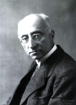
By Franz Keibel
The first relatively satisfactory synopsis of the development of the external form of the human body is that given by His[1] in his "Anatomie menschlicher Embryonen" and in the Normentafel published with it. In the latter there is shown a series of human embryos dating from the end of the second week to the end of the second month. With this latter period the development of the embryo is so far advanced that the human in it is recognizable even to the laity ; His designates this as the embryonic period and that succeeding it up to birth he terms the fetal period. A comprehensive account of the development of the body during the fetal period, with abundant illustrations, has been given by Gustav Retzius[2] in his memoir "Zur KenntAis der Entwicklung der Korperform des Menschen wahrend der fetalen Lebensstufen", published in 1904.
Disregarding studies of individual embryos there must also be mentioned here the "Normentafel zur Entwicklungsgeschichte" of Keibel and Elze,[3] Carl Rabl's "Entwicklung des Gesichtes"[4] and the splendid heliogravures of human embryos that Hochstetter[5] has published. Those who desire a comparison of human development with that of animals I would refer to Hertwig's "Handbuch"[6] in which I have considered the development of the external form in vertebrate embryos.
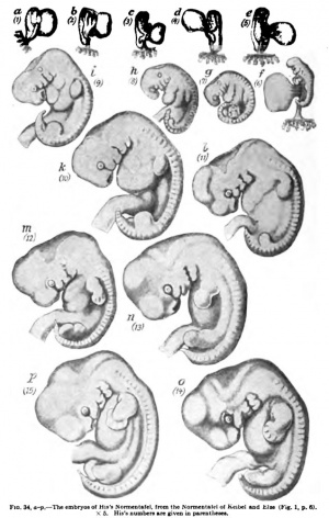
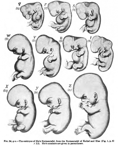
On account of the fundamental importance of His's "Anatomie menschlicher Embryonen," I here give a view of the development of human embryos as shown in His's Normentafel, the first fifteen figures, as in the original, being enlarged five times, the remaining ones only two and a half times.
|
|
Fig. 34. The embryos of His's Normentafel, from the Normentafel of Keibel and Elze (Fig. 1, p. S) X 2.B. His's numbers are given in parentheses. The individual embryos are lettered, His's numbers being given in parentheses. (See also full single figure)
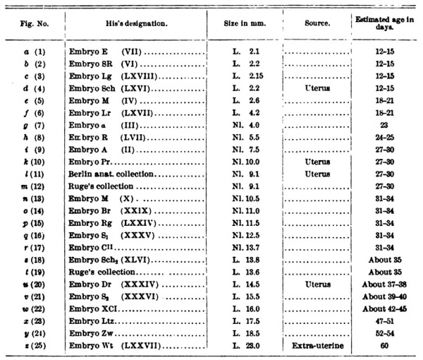
To this reproduction of His's Normentafel I append a synoptic table from which it may be seen how His designated each embryo, its size, and its age, as estimated by His.
To avoid repetition I shall not proceed to describe His's Normentafel, but will consider a series of embryos which, in my opinion, present the best summary now available of the development of the external form of the human body ; and in doing so I shall have occasion to consider the embryos of the Normentafel in their appropriate places. The so-called membranes will be described only in so far as they influence the form of the body.
Nothing need be added here concerning the youngest human embryonic disks to what has already been said in the chapter on "The Youngest Human Ova and Embryos." The embryonic disk of the Peters ovum had a length of 0.19 mm. ; whether it possessed a very small primitive streak must remain doubtful. In Spee's Von Herff ovum the disk had an oval outline and presented a median groove lying between the dorsally convex lateral portions, which were somewhat unequal in the transverse direction. This groove is the primitive groove, and it may be supposed that the primitive streak extends through the entire length of the embryonic disk, since Spec says: "The entire anlage of the embryonic disk is apparently only a portion of the actual primitive streak region."
Close upon the embryome disk of Spee's Von Herff ovum follows that of the Frassi ovum. In the former the primitive streak was at the height of its development, extending as it did throughout the entire germ; in the Frassi ovimi it is about half the length of the embryonic disk, and at its cranial end it shows the anlage of a neurenteric canal, while at the caudal end, as has been noted (p. 36), the anlage of a eloacal membrane has appeared. In front of the primitive streak there is a shallow medullary groove bounded by low medullary folds. The entire embryonic disk is slightly convex in the craniocaudal direction and from right to left, and it covers the yolk sack like a lid.
Following this embryo that of Spee's Glaevecke ovum (Fig. 37) may be mentioned. The actual embryo had a somewhat "constricted pear-shaped" outline, and within this the outline of the biscuit-shaped medullary plate was distinctly marked. The caudal end of the embryo was bent Snarpiy centrally, almost at a right angle, and consequently, when looked at from above, appears greatly foreshortened. SHglitly cranial to this downwardly bent portion there is a somewhat circular swelling, that in position corresponds to Hensen's node and surrounds, like a low wall, a roundish triangular opening, the neurenteric canal; behind this swelling is the primitive groove, resting upon the primitive streak. Anteriorly the primitive streak is embraced by the medullary folds. For the measurements of the embryo consult Chap. IV, p. 39.
Next to this embryo comes embryo 1 of His's Normentafel. This embryo was obtained from an ovum that ; measured in the fresh condition 81 X 5.5 mm. and was completely surrounded by villi. The length of the embryo, including the body stalk, was 2.6 mm, and without the body stalk was 2.1 mm. The yolk sack was somewhat flattened and measured 2.3 X 1.6 mm.
The embryo rested upon the yolk sack for about 2 mm. of its length and was enclosed in an amnion which also surrounded the upper surface of the belly stalk. In the embryo itself, the sectioning of which was unsuccessful, the medullary folds and medullary groove could be recognized, and laterally from the medullary folds at the anterior extremity the anlage of the heart was visible.
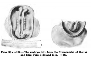
I place next the embryo Klb. of the Normentafel of Keibel and Elze, Figs. Hid and Ilia. The ovum which contained the embryo was obtained by a laparotomy and both it and the embryo may be regarded as quite normal. The embryo had five to six pairs of primitive somites. The vertex bend had already begun to form, otherwise the embryo lies flat on the yolk sack; it has no dorsal bend. Fig. 38 shows it from the dorsal surface, and, the amnion being cut away, it is seen to be attached to the chorion, a small portion of which is represented, by a short belly stalk. To the right and left of the cut edges of the amnion the yolk sack projects beyond the embryonic structures. Both the cranial and caudal ends of the embryo are separated from the yolk sack, but the middle portion is still spread out. The medullary groove is widely open, but rather deep. Caudally the medullary folds embrace the dorsal opening of the neurenteric canal and the cranial end of the primitive streak, which immediately bends downward so that it cannot be perceived in its entire extent; indeed, it lias already undergone much retrogression. The well-marked medullary anlage shows the vertex bend, so that its cranial end cannot be seen in a dorsal view. The brain part of the medullary anlage shows a separation into three portions, the most caudal of which extends to about the fourth pair of primitive somites and then passes without any marked boundary into the spinal cord. I may here note that by far the greater part of the dorsal region of this embryo belongs to the later head region. The boundary between the head and trunk regions passes through the fourth primitive somite. The most anterior somite of the neck region is first differentiated.
Fig. 39 shows the embryo from in front. The amnion has again been removed; ventrally the relatively large yolk sack is seen. Of the embrjonic anlage only the ventrally bent portion, as far back as about the vertex bend, can be seen; but it may be observed that the most anterior portion of the brain anlage is relatively greatly developed. The medullary groove terminates a little behind the cranial end of the medullary aniage, so that the latter shows anteriorly a transverse ridge. Nothing is yet to be seen of the optic pits, the forerunners of the optic vesicles.
Regarding the measurements of this embryo Kromer makes the following statements: "The greatest length of the embryonic aniage from the anterior amniotic border of the head cap to the chorionic end of the belly stalk was 1.95 mm. ; the length of the embryo, without the belly stalk, from head to tail was 1.8 mm. ; the greatest breadth of the yolk sack was barely 1.2 mm. ; the breadth of the embryonic disk at the junctioti of the amnion and yolk sack (measured in an anterior view) was 0.9 mm." The size of the yolk sack was 1.1 mm. iu height, 1.1 mm. in breadth, and 1.5 mm. in length.
After this embryo, that represented in Fig. 2 of His's Normentafel may follow, although on account of its possessing a dorsal flexure I am somewhat in doubt if it is normal in form. Its greatest length was 2.2 mm. The greatest diameter of the somewhat collapsed yolk sack was 1.9 mm.; and the embryo rests upon it in such a way that anteriorly the head end, for a distance of 0.4 mm., and posteriorly the caudal end, for a distance of 0.5 mm., project beyond the yolk sack navel, which is 1.3 mm. in length.
The body of the embryo shows the middle of the dorsal surface somewhat depressed and the head and tail ends are somewhat bent downwards. The margins of the medullary plate are still wide apart, and a number of primitive somites, how many could not be exactly determined, were formed. The aniage of the heart was still paired, and upon the surface of the yolk sack there were numerous wartlike elevations (anlagen of blood-vessels).
The Bulle embryo of Kollmann (Fig. 40) may well come in here or even after the embryo Klb. -20(Fromti* Both the head and tail ends project beyond the yolk Kei™'^*EiM, sack, which communicates widely with the intestine. Fig. iv, plw s.) BO that one cannot yet speak of a yolk stalk. The yolk sack has been cut away, except for the part by which it is continuous with the embryo; and similarly the amnion has been cat away not far from its root; under the caudal end of the embryo is to be seen the belly stalk. The figure shows the embryo from behind and from the right, so that the heart swelling is hidden. The brain portion of the medullary canal, in which the segments of the brain can be seen, is still open, and the caudal end of the canal is also open, although this cannot be perceived from the figure. If we reckon three of the fourteen primitive somites as belonging to the head and eight to the neck region, there still remain three as representing the thorax ; in the relatively small caudal end of the embryo almost all the segments of the trunk and tail must still be represented. In the region of the sixth to the fourteenth somites the dorsal surface of the embryo is slightly depressed. Kollmann says: "In the region of the sixth primitive somite the flexure of the dorsal region, which later becomes so striking, is noticeable." I cannot recognize in the figures of the embryo a flexure in the region of the sixth somite, and in any case the sixth segment belongs to the neck region. That a dorsal flexure occurs at this stage or later in normal human embryos I regard as disproved, and in this Spee[7] is in agreement with me. In 1905[8] I had come to the conclusion that even if a dorsal flexure normally occurs in human embryos it can only be at a stage of development in which six or at the most twelve primitive somites are present. I do not, however, regard the evidence of its occurrence at this stage as sufficiently based upon the embryo that Eternod[9] has had modelled by Ziegler and upon an embryo with seven ])airs of primitive somites belonging to Graf Spee.[10]
It is a question whether in this and other cases one has not to deal with a deformity produced by swelling. (For further consideration of this matter see the Normentafel of Keibel and Elze, pp. 22-23.)
The greatest length of the embryo, measured after it had been preserved in alcohol, was 2.36 mm. The embryo Pfannenstiel III (Figs. 41 and 42), figured in Keibel and Elze's Normentafel, has the same number of primitive somites as the Bulle embryo of Kollmann, yet it is more developed. It was obtained from a hysterectomy and was modelled by Elze. The medullary tube is open in the brain region as well as caudally; but, in addition to the vertex bend, present in the embryo Klb., a nape bend has appeared ; there is no indication of the dorsal flexure. The optic pits were distinct and the auditory epithelium was slightly depressed. The greatest length of the embryo measured 2.6 mm.
The flexure which I have termed the nape bend was very striking in this embryo. If it be normal, it is very doubtful if the nape bend that is found in later stages is comparable with it, and if it does not later disappear before the well-known nape bend of later stages develops. Older embryos of mammals and also of man, such as embryo 3 of His's Normentafel, still lack a nape bend, as does also an orang-outang corresponding to this last embryo.[11]
At this point a great gap unfortunately occurs ifi our aeries of human embryos, so that a settlement of the question is at present impossible. The embryo shown in Pig, VI of the Normentafel of Keibel and Elze had already twenty-three pairs of primitive somites ; in addition, our ideas as to its external form are based upon a not very suCoessful model, whose head region presents a very improbable form. I shall not, therefore, give a figure of the embryo here, but merely describe it briefly. It shows a well-marked vertex bend, but no distinct nape bend; and it is spirally coiled, so that its tail end comes to lie to the right of the very large belly stalk. The medullary tube is still open at its caudal end. The heart swelling lies entirely in the amniotic cavity, and through its thin wall the heart must certainly have been shadowed. Three branchial arches and as many branchial furrows were recognizable, and dorsal to the second furrow there was the already greatly narrowed opening of the ear vesicle. The extremities have not yet formed.
Embryos 3, 4, and 6 of His's Normentafel, Fig. 34, c (3), d. (4), and ^ (6) I regard as abnormal; c (3) and d (4) show the much discussed dorsal flexure and / (6) seems to me for other reasons to be excluded from the series.
The embryo shown in Fig. 34, e (5), seems to me more worthy of confidence; and I repeat it here, more highly magnified, as Fig. 43. It came from an ovum which measured 7.5-8 mm. in diameter and was completely surrounded with villi. The amnion lies close to the embryo and does not yet entirely enclose the heart. The body of the embryo is bent upon itself anteriorly and at the same time twisted about its axis so that the head end is turned toward the left and the pelvic end toward the right. The dorsal curvature is very regular, and the nape prominence is not yet pronounced. The anterior portion of the head is bent ventrally to such an extent that the vertex is formed by the midbrain. Behind the anterior part of the head there is a deep depression, which indicates the entrance of the oral sinus and is continued dorsally in the orbitonasal groove. Behind the mouth cleft is a broad mandibular process, separated by a groove from the second visceral arch; and the posterior boundary of this second arch can also be made out. The study of the entire embryo revealed no clear picture of the third and fourth arches, although their presence was quite evident in sections. The anlage of the heart projected from the ventral surface of the body as a broad transverse swelling; its prolongation on the right side extends forward as the aortic bulb to the edge of the mandibular process. To the atrial portion of the heart belongs an outpouching which is seen ventral to the hinder portion of the head, on the lateral wall. Immediately behind the heart the umbilical vesicle (yolk sack) projects from the umbilicus, which has the form of a longitudinal cleft; the yolk sack was somewhat sunken in and was pyriform in shape.
The pelvic end of the body is curved forward in a hook-like manner and on account of the twisting of the embyro cannot be seen from the left side. In the posterior half of the trunk one sees four parallel longitudinal ridges, two of which, the medullary and somitic ridges, belong to the axial zone, while the other two, the Wolffian and marginal ridges, belong to the parietal zone. No traces of the extremities are yet visible. According to His's estimate there were thirty-five pairs of primitive somites. His gives the following measurements:
- Greatest length in a straight line 2.6 mm.
- From vertex to behind the mandibular process 0.7 mm.
- From vertex to behind the heart 1.4 mm.
- Height of yolk sack where it projects from umbilicus 0.6 mm.
- Maximal height of yolk sack 1.7 mm.
- Length of yolk sack 2.6 mm.
- Length of the posterior portion of the body, measured from the point of emergence of the yolk sack 0.6 mm.
The first embryo of Hochstetter's series (Fig. 44), which belongs to Professor Fischel of Prague, presents a very great advance in development. As the figure shows, it is not only very greatly curved upon itself, but it is also strongly twisted spiraUy. In the head one sees the primary optic vesicles and on the first branchial arch an early indication of the maxillary process. Both upper and lower extremities are recognizable, the former being plate-iike. The definitive nape bend is strongly developed. The greatest length of the embryo was 4.02 mm; it was obtained as an abortion.
The embryo which I would place next in the series is embryo G 31 of the Anatomical-biological Institute of Berlin (Fig. 45) {Fig. VIII of the Normentafel of Keibel and Elze) ; it stands close to embryo 7 (Fig. 34, g) of His's Normentafel, Its greatest length is the nape-breech length (nape line) and is 4.9 mm.; the vertexbreech length is 4.7 mm. It is rather strongly curved upon itself, but more regularly than the preceding embryo ; the nape bend is strongly marked, and the spiral twisting is still quite distinct. The tail lies to the right. The upper extremities are plate-like in form, the lower ones ridge-like. The anterior end of the head shows in the middle line a slight depression, and the anlagen of the eyes show through the integument. The region of the later sinus cervicalis is still but little depressed, so that the third and fourth branchial arches are quite easily seen. The atrial and ventricular portions of the heart can be distinguished through the thin wall of the pericardial cavity.
A few words may be said concerning embryo 7 of His's Normentafel(Fig.34,5). This embryo has reached the highest degree of curvature that has yet been observed in the human embryo. The length from the forehead to the tip of the coccyx following the curvature was 13.7 mm.; the straight line from the nape prominence to the twelfth thoracic segment, the nape line (NL), was 4 mm. in length. The tail end was curved forward to the level of the ventricle of the heart and lay upon the left side of this. In the curve which is formed by the embryo from the forehead to the tip of the coccyx there are four regions where the bending is stronger than elsewhere: (1) the region of the mid-brain; (2) the nape prominence; (3) the boundary between the neck and thoracic regions; and (4) the boundary between the abdominal and pelvic regions. The plane of symmetry of the embryo is warped and is twisted in such a way that the head looks to the right and pelvic end to the left. The upper extremities are plate-like, the lower ridge-like.
At the head end the shape of the cerebral hemispheres, the tween-brain, mid-brain, hind-brain, and after-brain, is plainly recognizable, and the boundaries of the fourth ventricle are sharj)Iy defined. In a view from the dorsal surface the lateral walls of the fourth ventricle show a series of regular and bilaterally symmetrical transverse folds (neuromeres). The optic vesicles form on each side a circular circumscribed projection measuring 0.35 mm. in diameter, and the auditory vesicles are very evident as oval structures lying at the level of the second visceral groove. In addition to the moderately developed maxillary and mandibular processes the lateral wall of the bead shows on each side three visceral arches, the fourth of which is still fully exposed. The distance from the anterior border of the ma-xillar}process to the fourth branchial groove was 1.4 mm,; a line drawn through the ventral ends of all four arches was almost straight and cut the fore-brain some distance in front of the optic vesicles. The anterior borders of these were about 0.55 mm. from the anterior pole of the fore-brain, so that the prominence of the fore-brain characteristic of human embryos in all later stages of development is already present. Immediately behind the ends of the visceral arches is the heart, the three portions of which are plainly visible when the embryo is examined from both sides, with this difference, however, that the atrial swelling is more distinct on the left side and the swelling of the aortic bulb on the right side. No deep cleft yet separates the swelling produced by the aortic bulb from the facial surface of the head. The visceral region of the body lying behind the heart shows on the right side a distinct Wolffian ridge and the transition border of the amnion; on the left side no characteristic elevations are visible.
Near Fig. 8 of His's Normantafel (Fig. 34, h) stands the embryo shown in Fig. 46. Jt was obtained as an artificial abortion. It shows the vertex and nape prominences and is not only strongly curved on itself but is also spirally twisted. The olfactory area is beginning to show a better delimitation dorsaily and laterally, and the maxillary process can be seen in a profile view. In the region of the sinus cervicalis which is still widely open, the third and fourth branchial arches are plainly exposed; the heart swelling is very prominent and the atrial and ventricular portions of the heart are recognizable. The liver swelling la still but poorly developed, and the tail is curved in between the heart swelling and the belly stalk. Dorsal to the primitive somites ore sees O. Schultze's[12] division of the sclerotomes. The embryo had thirty-six pairs of somites.
Between embryos 8 and 9 of His's Normantafel (Fig. 34, h and i) there is a rather wide interval, into which the embryos shown in Fig. X and Fig. XI of the Keibel-Elze Normentafel fit. The erabryo shown in Figs. XII and XIr corresponds fairly well with the Brausembryo shown in Figs. 2 and 3 of Hochstetter'S Fig. 47. — The Brsm embryo. XIO. (From Hochstetter's series. For explanation of the figure of this, shown in Fig. 47, a few words will sufiice. The olfactory area is beginning to be delimited more sharply dorsaily and caudally; the first and second branchial arches have now become relatively large and have begun to be further modified; the triangular area caudal to them, in which the third and fourtli arches are situated, is slightly depressed, forming the sinus cervicalis; the upper extremities have passed from the plate-like into the stump-like stage.
Following closely upon this embryo comes embryo 9 of His's Normentafel (Fig. 34, i). In it the nape bend is more strongly marked, so that the head is much more bent down upon the heart; the distinctly circumscribed olfactory pit rests upon the heart. The tips of the anterior extremities have become bent ventrally, and the roots of the extremities are correspondingly angled. In the trunk one sees three tumor-like projections, of which the two situated more cranially and ventrally are produced by the ventricular and atrial portions of the heart, while that lying more caudally and dorsally is formed by the liver. The embryo shows a very distinct external tail. The external configuration of the head is principally determined by the subdivisions of the brain, whose shapes are clearly recognizable through their thin covering. At the base of the fore-brain is the olfactory pit, and a slight distance in front of this is the eye with the lens groove. The smallness of the eye oomi)ared with the great development of the forebrain is characteristic. A prominence behind the eye marks the position of the ganglion of the trigeminus; it lies in the angle between the raid-brain and hind-brain. At the level of the second visceral groove the auditory vesicle, together with the ganglion acnsticum lying in front of it. forms a slight elevation. Elevations are beginning to appear on the mandibular process and hyoid arch ; the third and fourth branchial grooves lie at the bottom of a triangular depression, which will become the sinus cervicalis.
Embryo 10 of His's Normentafel is comparatively large for the degree of development it presents; it resembles the preceding embryo, and was taken from a uterus.
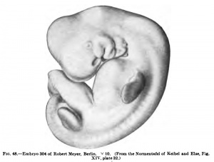
Embryo XIV of the Keibel-Etze Normentafel (Fig. 48) is not quite so large, but is more developed. This is shown especially by the condition of the extremities and of the sinus cervicalis. Behind the dorsal part of the hyoid aroh one may perceive the entrance into the glossopharyngeal organ (the second branchial groove organ). In the upper extremities the band plates are distinct Forty trunk somites were counted, and the last six of these were beginning to be transformed into a tail filament. The embryo was obtained from an artificial abortion.
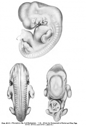
| The embryo to which we now come may be considered not only from the left side (Fig. 49) hut also from the dorsal (Fig. 50) and ventral (Fig. 51) surfaces. It is the Hoehstetter embryo Ma. 3, Fig. XVII of the Keibel-Elze Normentafel and Figs. 7, 8, and 9 of Hochstetter's series. The trunk region has again begun to elongate. The spiral twisting is evident only to a slight extent in the tail region, the tail lying to the left of the belly stalk. The nape bend is almost a right angle; the cerebral hemispheres are recognizable externally; and the cerebellum shows out plainly, especially in the ventral view (Fig. 51). The axes of the upper limbs are almost parallel to the dorsal line; the hand plates are almost circular; the elbows show especially distinctly in the dorsal view (Fig. 50); and in the same view one may perceive, on the surface of the upper limb which looks towards the trunk, a small tubercle. In the lower limb the foot plates are marked off. Dorsal to the row of primitive somites one can clearly distinguish Schultze's segmentation of the sclerotomes. The maxillary processes have come into relation with the median nasal process, the openings of the nasal pits look towards the wall of the pericardium and are no longer to be seen from the side, especially from the left. A distinct sculpturing is present on the mandibular process and especially on the hyoid arch. The entrance into the sinus cervicalis is still visible as a small triangular hole. |
In part somewhat more and in part less developed tlian this are embryos 11, 12, and 13 of His's Normentafel (Fig. 34, I, m, and n) ; that shown in Fig. 11 (I) was taken directly from the uterus. In that shown in Fig. 12 (m) a small nape furrow has formed beneath the strong nape prominence and the position of the upper extremities does not seem normal. In that shown in Fig. 13 (n) the body is relatively slender and a heart swelling cannot be made out.
The body of Hochstetter's embryo P. 1 (Fig. 52) (No. 10 of Hochstetter's series, Fig. XIX of the Normentafel of Keibel and Elze) is already rather elongated. The elbows have moved out from the body and the forearm makes an acute angle with the dorsal line. The fingers are beginning to become distinct on the hand plates, and tlje foot plates have become circular. Close to this embryo come those shown in Figs. 14, 15, 16, and 17 of His's Normentafel (Fig. 34, o, p, q, and r) ; in Figs. 15, 16, and 17 the thumbs are distinctly recognizable.
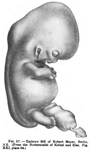
In the lower limbs of Figs. 18, 19, and 20 of His's Normentafel (Fig. 34, 5, t, u, and v) the aniagen of the toes can be seen. The head is gradually bending to the erect position, and in correspondence with this the neck is forming; the formation of the external ear is also making progress. These stages may be represented here by Hochstetter's embryo Chr. 2 (Fig. 53, No. 15 of Hochstetter's series, Fig. XX of the Keibel-Elze Normentafel), Eabl's embryo C (Figs. 54, 55, and 56, Nos. 16, 17, and 18 of Hochstetter's series) and the embryo No. 302 of Robert Meyer's collection (Fig. 57, Fig. XXI of the Normentafel of Keibel and Elze).
In the embryo Chr. 2 the nape bend is somewhat more than a right angle. The mouth lies in close contact with the pericardium, and the nose has become free from it. The folds of the pinna, the tragus and the antitragus, are formed; the concha is widely open and flat. Behind the nape prominence the contour of the back shows a distinct depression. The fingers have become quite pronounced and the toes are just indicated, but the position of the great toe is already to be recognized. The elbows and knees are evident, and the angle that the axis of the forearm makes with the dorsal line is almost a right angle.
Fig. 54-56.
Slightly further developed than this is Rabl's embryo C (Figs. 54, 55, and 56). The nose is more marked off, the eyelids are further advanced, and so also the ear. The finger-tips are distinctly projecting beyond the border of the hand plate. In the ventral view one may note the position of the eyes and of the limbs. Between the foot plates may be seen the physiological umbilical hernia. The tail has become transformed into a coccygeal tubercle. In the dorsal view the sharp slope of the caudal portion of the trunk is striking.
Fig. 57.
The embryo 302 of Robert Meyer's collection (Fig. 57) shows a distinct depression between the root of the nose and the forehead. The pinna, tragus, and antitragus are evident; and while the concha is still widely open it shows a tendency to deepen. Early indications of the eyelids are to be seen. The nape flexure is still very evident, but it forms an obtuse angle, and the depression of the dorsal line just below it is less marked. The nose and mouth have both separated from contact with the pericardium ; the mouth is slightly open and the mandible lies close upon the breast. The shoulder is distinctly marked off from the body, the upper arm makes an angle with the forearm, and the elbow projects strongly. The hand plate is beginning to turn ventrally, and the finger anlagen are very distinct; their tips are becoming free and the thumb is strongly abducted. In the lower limb the knee is very distinct and anlagen of the toes are appearing on the foot plate. The dorsum of the foot is not yet marked off from the crus.
When we pass from embryo 21 (Fig. 34, v) to embryo 22 (Fig. 34, w) of His's Normentafel, we are passing from the embryonic to the fetal stage. The profile of the latter embryo already shows clearly a nose, upper lip, lower lip, and chin ; and the neck is also present The eyelids are formed, and above the eyes there is a distinct supra-orbital swelling. The upper limb, in which tho fingers have become distinct, has increased in length considerably and shows a distinct division into upper arm, forearm, and hand. The shoulder has formed, and the characteristic position of the arms is worthy of note. In the lower limb the foot, crus, and thigh can be distinguished. The aniagen of the toes are not yet separated from one another, but that of the great toe ia especially well marked. The nape prominence and nape depression are much reduced, and the coccygeal tubercle still appears as a short stumpy tail.
With the three remaining stages of His's Normentafel, Figs. 23, 24, and 25 (Fig. 34, x, y, and z) the transition from embryo to fetus has been accomplished. In Fig. 34, x, one sees the toes separated from each oUier, and the great toe has a characteristic abducted positiou, recalling the position of the thumb in the corresponding stage in the development of the hand. In Fig. 34, y, the foot is better formed ; the legs have undergone a twisting so that the knee looks more upwards and the foot more downwards. Fig. 34, e, still shows a slight nape prominence and a very shallow nape depression. The small creature shows a distinctly human character in its features. Some additional figures may complete the story.
The embryo shown from in front in Fig. 58 corresponds somewhat to Fig. 22 {Fig. 34, w) of His's Normentafel. The position of the eyes, the broad nose, and the position of the limbs may be noted.
A fetus measuring 25 mm. in its greatest length js shown from the left side and from the ventral aspect in Figs. 59 and 60, reproduced from the Normentafel of Keibel and Elze. It may be regarded as standing between Figs. 24 and 25 (Fig. 34, y and z) of His's Normentafel. Again I would call attention to the position of the limbs. In Fig. 59 we see touch pads on the sole of the right foot. At the summit of the coccygeal tubercle there is a small knob, as in all well-preserved embryos of this stage, and it is also seen in the ventral view (Fig. 60), in which the physiological umbilical hernia is indicated by the coils of the intestine showing through the wall of the cord.
We may now again pass in review the stages which the human embryo must traverse in order to acquire its human form. The earliest known embryos are flat, shield-like plates which rest upon the yolk sack. At a certain stage a primitive streak extends throughout the entire length of the shield; in front of the primitive streak the embryo is formed, and it grows at the expense of the streak, which retrogresses in a craniocaudal direction. The streak is thus converted into a growth zone, which may be termed at first the trunk bud and later the tail bud. To correctly understand these processes of development we must bear in mind the extent to wliich the head region predominates in young embryos.
Beturning again to the primitive streak, during its modification its caudal end becomes bent ventrally; the cloacal membrane had previously formed in this caudal region, and it now comes to lie on the ventral surface. This bending process is associated with the constriction of the embryo from the yolk sack ; cranially and caudally, from the right and from the left, the boundary grooves cut inwards and soon convert the yolk sack into a stalked vesicle; in the meantime the dorsal portion of the embryo grows more rapidly than the ventral, a condition caused by the accelerated growth of the medullary anlage. Before the medullary plate is converted into a tube the brain, with its principal subdivisions, becomes distinct at the anterior end, and the optic pits are also formed. By the rapid growth of the brain portion, while the medullary tube is still open, there is produced a bending down of its cranial portion (the vertex bend) and, if the Pfannenstiel III embryo {Figs. 41 and 42) is to be regarded as normal, also the more caudal nape bend. It has been already pointed out, however, on pp. G6 and 67, that a difficulty exists in this particular. Embryos that are properly believed to be older do not show the nape bend distinctly, and these agree with the embryos of other mammals, especially with that of the orang-outang which I described in. 1906. There are two possibilities : either the nape bend of the embryo shown in Figs. 41 and 42 was an abnormality; or it is a primary nape bend that normally disappears, the long known nape bend later appearing independently of it. On account of the lack of the necessary stages this question cannot be decided at present; but in any event the entire embryo soon becomes curved on account of the much greater growth of the dorsal surface, and coils itself in a spiral. The spiral turns sometimes to the right and sometimes to the left. While these developmental processes are taking place there has formed from the tail bud a small but unquestionable external tail. In these stages also the head region still predominates to an extraordinary degree, but the embryo no longer consists, as in the stages of the early primitive somites, only of the future head, so to speak. The extensive coiling toward the ventral surface is followed by a very gradual uncoiling; first the trunk straightens out and the nape bend slowly becomes obliterated. These processes are accompanied, perhaps determined, by a rapid growth of the ventrally situated organs, especially of the heart and liver, which produce on the surface of the body the heart and liver prominences. The Wolffian bodies do not play a very important part in this respect in man. With the straightening out of the nape bend opportunity is afforded for the formation of the neck. Originally the face lies closely upon the heart prominence and the ventral portion of the neck is entirely wanting, "While its lateral parts are occupied by the branchial or visceral arches. The transformation of these interesting structures will be thoroughly described in other places; here the following may suflSce. The third and fourth arches — quite transitorily there is also recognizable the aniage of a fifth — remain small in comparison with the first and second, the mandibular and hyoid arches, and consequently the region in which they occur becomes depressed, forming the sinus cervicalis, which becomes covered in by the second or hyoid arch and its opercular process. In the region of the first branchial groove there are formed on the mandibular and hyoid arches a number of elevations and folds from which the external ear is formed; its development will be considered in detail with the sense organs.
In determining the form of the head the brain is at first almost the controlling organ, and its various parts can be distinguished until relatively late stages through the thin walls of the head. Gradually the mesenchyme increases in the walls, and, above all, the face begins to develop in the service of the principal sense-organs and of the respiratory and digestive tracts; its formation will be considered later on.
The ventral wall of the trunk is at first very thin, and the heart with its various parts, the liver, and other viscera may be seen through it. From the right and left the anlagen of the skeleton and the musculature grow into the walls; inhibitions of this growth may occur and produce ectopia cordis and fissura stemi, which depend upon disturbances of this process in the thoracic region. A portion of the intestine normally projects like a hernia into the umbilical cord, in association with the outward growth of the abdominal walls; thus the hernia funiculi umbilicalis physiologica is produced. Normally this hernia disappears with the further formation of the abdominal walls, but occasionally it may persist as an inhibition structure.
On the ventral wall of the body the region from the umbilicus to the end of the trunk is the original primitive streak territory. That portion of the streak which was originally, before it became bent ventrally, the caudal portion, but which is now directed cranially, becomes encroached upon by the developing skeletal and muscle anlagen of the trunk. If inhibitions occur in this region ectopia vesicae and pelvic clefts may be produced, and epispadias also belongs in this category. The openings of the urogenital sinus and the anus and the intervening perineum arise in the territory of the portion of the cloacal membrane which persists after the formation of the abdominal wall below the umbilicus. For an account of these developmental processes reference may be had to the chapter on the urogenital apparatus.
That a tail occurs in the human embryo has already been noted; embryos of 4-12 mm. NL. have a typical external tail, in which a caudal gut occurs and which possesses more segments and spinal ganglia than persist. This external tail (cauda aperta) becomes transformed in its distal portion into a tail filament, which is knob-like in the human embryo and is later cast off, while the remaining portions become overgrown by the neighboring parts and so disappear beneath the surface; in this situation remains of it may persist, forming what is termed the internal tail (Braun, 1882) or cauda occulta (Rodenacker, 1898).[13]
If one now glances at the body as a whole, one can hardly fail to be struck by the fact that the cranial portion is much more fully developed, up to relatively later stages, than the caudal portion ; this is shown very clearly by the dorsal views of embryos, and I would call attention, in this connection, to Figs. 51 and 55.
The extremities first appear as ridges which later are converted into plates, becoming more sharply defined cranially and caudally. The portion which is first formed corresponds essentially to the anlage of the hand or foot ; gradually the anlagen of the forearm and crus, and then those of the upper arm and thigh, grow out from the body. The further differentiation of the extremities, as well as tliat of the face and head, will now be considered more thoroughly, and for this purpose good figures are much more important than extensive descriptions.
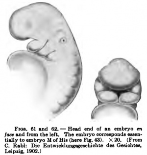
The figures showing the development of the face in the first stages of its formation are rather few in number, since the processes cannot be well observed in the human embryo without dissection, and dissection prevents the obtaining of a continuous series of sections. I shall start with a stage that Rabl estimates at nineteen to twenty days ; According to the estimate of Bryce and Teacher (see pp. 26 and 27) it would be considerably older. It corresponds essentially to Fig. VI of the Normentafel of Keibel and Elze and to embryo 5 (Fig. 34, e) of the His Normentafel. Fig. 61 shows a profile view of it. Three branchial arches are recognizable ; on the first the maxillary process cannot be seen in this view, although it is present, as may be seen from the front view (Fig. 62). The optic vesicles, which are directed laterally, show through the wall of the head, and over the second arch are the auditory vesicles, not yet quite closed. In the front view (Fig. 62) one looks into the pentagonal oral sinus ; it is bounded above by the frontal process, laterally and above by the maxillary processes, and below by the mandibular processes. Much more developed is the face that I reproduce (also from Rabl) in Fig. 63. It is from an embryo which measured 8.3 mm. in the preserved condition and belonged, According to Rabl's estimate, to the fourth or perhaps the beginning of the fifth week. It corresponds fairly well with Fig. 11 .
(Fig. 34, l) of His's Normentafel and to Fig. XIV of those of Keibel and Elze. The pentagonal opening of the oral sinus has transformed into a broad mouth-cleft, whose lower border shows a notch between the two adjacent ends of the mandibular processes. The nasal pits are distinct; they are deeper dorsaliy and are flattened out ventrally. One may also speak of the two nasal processes, that is to say, the ridges which bound the nasal pits laterally and medially. The portion of the frontal process between the medial nasal processes is still very broad.
The advance that is to be seen in Fig. 64 (again from Rabl) is quite marked. The figure represents the face of an embryo that corresponds to Fig. 14 (Fig. 34, o) of His's Normentafel and to Fig. XIX of those of Keibel and Elze; its napebreech length was 11.3 mm. and its age was estimated by RabI at thirty to tliirty-one days. In the head the fore-brain region is markedly prominent; below the forehead the area triangularis (His) projects, and below the middle of this there runs what is only a moderately broad groove through the mouth opening into the palate, separating the medial nasal processes, which at their first appearance were so far apart. The two nasal openings are not only relatively but absolutely nearer each other than in the preceding stage, and look directly forward. The lower ends of the medial nasal processes, which are uniting with the maxillary processes, are separated by a slight depression from the lateral processes and are the proccssus globularcs (His). The lateral nasal processes are consequently excluded from the formation of the upper boundary of the mouth, which becomes the upper lip.
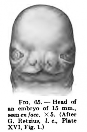
Fig. 65 shows the face of a 15 mm. embryo from in front, after Retzius. The upper lip is still divided, the nasal septum is still somewhat incomplete, and the processus glohulares are still visible. From the upper angle of the nose two folds run from above and medially downward and laterally, the medial one passing to the angle of the month; these are the oblique -maxillary folds, that is to say, the nasolabial and the suborbital folds. The eyes are directed distinctly laterally and are comparatively wide apart ; the mouth is very wide ; and at the middle of the lower jaw the anlage of the chin is visible, although the lower jaw and chin are as yet very poorly developed.
Figs. 66 and 67 show the head of an embryo of 18 mm. en face and in profile (after Retzius). The view en face appears rather remarkable; Retzius thinks that the embrj'o may not have been quite normal, but gives no further reason for such a supposition. The description given of the preceding embryo will answer for this one also, if it be added that the nasal septum is now completely formed. In the profile view the position of the ear should be noticed; if the mouth cleft be produced dorsally the ear would He below it. Figs. 68 and 69 show profile and en face views of the head of a fetus of about eiglit weeks and 25 mm. in length (after Retzius). The eyelids are in process of formation and supra-orbital folds aJso occur, in addition to the nasolabial and suborbital. The supranasal groove is strongly marked. The distance between the eyes is still considerable, and they are distinctly directed laterally.
Figs. 70 and 71 represent the head of a fetus 42.5 mm. in its greatest length (ninth week), in profile and en face. In the profile view the great development of the forehead region is striking, and below this the root of the nose is deeply depressed. The nose is still low, aeeaeniaa. xs. (After but the lower jaw and chin are well marked. The eyes are completely closed by the lids, but the distance between the inner angles of the eyelids is very considerable; from medially and above the interpalpebral fissures are directed laterally and downward. The upper eyelid is relatively small and is bounded above by a aharply-marked arched groove and below by the interpalpebral fissure; from the inner angle of the eye a supra-orbital groove extends obliquely laterally and upward. The nose is ven,' broad in proportion to its height (172:100), and the external nares are closed by the epidermal plugs which are continuous with an epidermal thickening on the upper lip.
Finally, the profile view of the head of a fetus 117 mm. in length may be shown {Fig. 72), and in it I would draw especial attention to the projecting upper lip and the receding chin, to the double lip, and to the shape of the nose. The pinna has almost the position it holds in the adult. Witii regard to the mouth it may be observed that the vertical median furrow which becomes the philtrum appears in the fourth month. The wall-like projecting margin of the upper lip is separated from the inner portion, which in the fourth and fifth months shows more or less projecting tubercle-like elevations. The middle portion of the margin early becomes the tuberculum labii superioris. In a similar manner the wait-like margin of the lower lip becomes delimited from the inner portion. In the first half of the third mouth the two lips project about equally, but later the border of the upper lip and the lip itself grow more rapidly, so that in the fourth and fifth months it projects markedly beyond the lower lip; by a stronger growth of the lower jaw and lip this difference is gradually overcome in the sixth to the ninth months, but by a kind of inhibition process the early fetal arrangement may be retained in the adult to a marked degree. Retzius has given especial attention to the time of occurrence of individuality, and comes to the conclusion that in man it is recognisable even in the fourth mouth of uterine life, and becomes more marked in the succeeding months.
The first phases of the development of the extremities up to the formation of the fingers and toes have already been considered in connection with the development of the entire embryo; an account of the further changes in the hand and foot may now be presented, the descriptions given by Eetzius being followed closely.
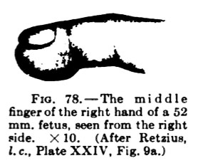
The hands, after the fingers are formed, early assume their human form and even in the third month have acquired their most important characters; they are then comparatively broad and the fingers are flexed. Of the persistent palmar furrows only the largest, the lines of Venus and Mars, are distinct in the third month. Four distal metacarpal pads (touch pads) appear at the beginning of the third month opposite the interdigital clefts, and in the course of this month they develop into strongly marked elevations. In addition, there is a distinct ulnar marginal pad on the metacarpus and one or sometimes two carpal pads. On the terminal phalanges strongly prominent, hemispherical touch pads develop. These, as well as the metacarpal pads, become relatively lower in the fourth and fifth months, and their boundaries become gradually more indistinct; only in individual eases do they persist in amore evident form with the later half of the fetal period. The feet are always somewhat behind the hands in their development. Even long before the separation of the individual toes by the interdigital clefts the abducted position of the great toe is striking; and very early, even in the second month, the prominence of the heel is recognizable. The great toe is from the beginning somewhat thicker and the little toe somewhat smaller than the other tliree. The soles, like the palms, are at first directed medially, and consequently are opposed to one another. The dorsum of the foot is relatively very high in the third month, and also broad towards the roots of the toes. Compared with their position in the adult the feet as a whole have now an " oblique " position (the varoequinus position); the arch of the sole is beginning to develop.
During this time, at the beginning of the third month, a row of distal metatarsal pads appears as four or five roundish or oval elevations, and one soon sees clearly that they lie opposite the interdigital clefts ; it seems as though a shifting toward the lateral (fibular) side occurred. In the next stage, that is to say,in the latter half of the third month, there are four metatarsal pads which correspond to the interdigital clefts, the fifth seems to have shifted proximally on the lateral border of the foot. At this time the four first-mentioned pads are relatively at their highest stage of development, and simultaneously the pads on the terminal phalanges have developed to liemispherical plantar elevations. In the fourth and fifth months the distal metatarsal pads undergo a relative retrogre.ssion and their outlines become gradually indistinct; the phalangeal pads remain well marked, although their outlines become less pronounced.
Some figures may make these points clear. Figs. 73-76 represent the extremities of a fetus of 25 mm. Fig. 73 shows the right hand from the volar surface with the touch pads ; Fig. 74, the two lower extremities seen from behind and dorsally. The feet are seen from their fibular borders, and their plantar surfaces are turned toward one another. The high dorsum and the malleolar eminences should be noted. Figs. 75 and 76 show the right foot from the dorsal and plantar surfaces; the abducted position of the great toe and the metatarsal touch pads cannot be overlooked.
|
Fig. 77. shows the sole of the right foot of a fetus of 44 mm. with its touch pads |
Fig. 78. represents the middle finger of a fetus of 52 mm. from the side. |
In the descriptions of the development of the face, hands, and feet the conditions in the fetal period have already been considered. For the development of the form of the rest of the body reference must be made to GustavRetzius (l. c). Retzius has been the first to study thoroughly the proportions of the human body during the fetal period, and a reaumi of his most important results may bring this chapter to a close.[14]
He finds as follows :
- The entire body length, measured from vertex to heel, increases during the fetal period to a greater extent than the vertex-breeeh length, that is to say, the lower limbs continually increase in length.
- The relation of the height of the head to the vertex-breeeh length, gradually diminishes.
- A comparison of the length of the entire vertebral colmnn with the height of the head and with the different regions of the column shows that:
- The height of the head diminishes relatively to the length of the vertebral column.
- In general, only a slight change can be observed in the ratio of the cervical vertebrae to the entire column, although there is a certain tendency towards a relative shortening of the cervical vertebrte in the earlier stages.
- Scarcely any change, apart from individual variations, can be seen in the relation of the thoracic vertebne to the entire column.
- The relation of the lumbar vertebne to the entire column shows no appreciable change.
- The relation of the sacrococcygeal vertebrae to the entire Column also shows no material change; individual variations are, however, especially great, and then difficulties in the way of making exact measurements are worthy of note.
- The relation of the circumference of the head to the body length diminishes from the earlier stages.
- As regards the relation of arm length to body length, it was found that during the seeond and third months the arm grows to snch an extent that often even in the third month, and more certainly in the fourth and beginning of the fifth, it reaches its greatest relative length for the fetal period, its first maximum (37-42 per cent, of the body length).
- As regards the relation of the upper extremity to the lower and of the leg length to the body length, it may be said that the lower limb grows more slowly than the arm during the fetal period; at the end of that i>eriod it is scarcely as long as the arm, but after birth it soon surpasses it. The relative maximum of length for the fetal period (36-39 per cent, of the body length) is acquired by the leg at about the fifth month.
- A comparison of the arm length with the lengths of the upper arm, forearm, and hand shows that:
- During the period from the third to the tenth month the upper arm is about 39-42 per cent, of the entire arm length.
- In the relation of the arm length and that of the entire distal part of the arm (foreami and hand) there are no perceptible changes either of progression or regression during the fetal period.
- Also the arm length compared with the forearm length (without the hand), and
- The arm length compared with the hand length show no noteworthy changes of proportion dunng the fetal period.
- The relations of the length of the lower limb to those of the three portions of which it is composed may be stated as follows:
- The relation of the thigh length to the leg length shows no noteworthy changes from the third to the tenth month.
- In the relation between the leg length and that of the crus (omitting the height of the foot), a slight relative elongation of the crus is evident about or before the middle of the fetal period.
- In the relation of the length of the foot to the leg length there is a definite relative elongation of the foot, especially from the sixth to the eighth month.
- In the relation of the breadth of the iliac crests to the body length no actual change occurs during the fetal period from the third to the tenth month.
- As regards the proportions of the head and face during the fetal period, Retzius' results are as follows:
- Ratio of head length to head width: In the first months, while the cerebral hemispheres are still developing posteriorly, no measurements can be obtained that allow satisfactory comparison. Notwithstanding that Retzius worked with Swedish embryos and that the Swedes are a typically dolichocephalic race, it seems that there is a strong tendency to brachycephalism and, indeed, to a quite high degree of it.
- Ratio of head length to head height : This index is very high in the early months (112.5, 111.1, 100, etc.); it is still high in the third month (108.5, 104.2, etc.); but toward the end of this month it sinks (86.0, 81.6), and remains at about the same level from the fourth to the seventh months, with only a few individual variations upwards. If the figures given above are interpreted According to the standard employed for adult skulls they all denote hypsicephalism (75.1 and over).
- Ratio of head length to head circumference: In general, the circumference of the head is about or almost three times as great as the length. The index, which at first is smaller, increases during the third month to this value and remains about the same, with indiridual variations, to the seventh month.
- Ratio of head width to head height : This index shows a definite tendency to diminish, apart from individual variations.
- Ratio of head circumference to face height: The figures show no regular change until the seventh month; it is remarkable that during these stages iilmost the same values recur.
- ↑ His W. Anatomie Menschliche Embryonen I - Embryonen des ersten monats (Anatomy of human embryos - Embryos of the first month). (1880) Leipzig.His W. Anatomie Menschliche Embryonen II - Gestalt und grossentwicklung bis zum schluss des 2 monats (Anatomy of human embryos - Shape and general development to the closing of the 2nd month). (1882) Leipzig.
- ↑ Gustav Retzius: Biologiscbe Untersuchungen, neue Folge xi, 1904.
- ↑ Keibel and Elze: Normentafel zur Entwicklungsgeschichte des Menschen, Jena, 1908.
- ↑ Carl Rabl : Die Entwicklung des Gtesichtes, Leipzig, 1902.
- ↑ F. Hochstetter: Bilder der ausseren Korperform einiger menschlicher Embryonen ans den beiden ersten Monaten der Entwicklung, Munich, 1907 (published by F. Bruckmann).
- ↑ F. Keibel: Die Entwicklung der ausseren Korperformen der Wirbeltierembryonen, etc., Hertwig's Handbuch, 1906, vol. i, chap. 6 (published 1902).
- ↑ Schwalbe's Jahresbericht, Jena, 1906, neue Folge, toI. xi, 2 Abt., p. 225 (literaltire of 1905).
- ↑ Keibel: Zur Embrjologie des Menschen, der AfEen und der Halbaffen, Verh. Anat. Ges. (Genf), 1305; also in C. R. Soc. dea Anatomistes, 1905.
- ↑ Compare A. C. F. Eternod : II y a un canal notochordal dans I'embryon humajn, Anat. Anz., vol. svi, 1899.
- ↑ Graf Spee: Mitteilungen iiber den Verein selileswig-liolsteiniselier Aerzte, vol. xi, article 8, 1887.
- ↑ See F. Keibel: in Selenka's HenschenaiTen, 9 Lieferung, 1906.
- ↑ 0. Schultze: Ueber etnbryonale und bleibende Segment ienmg, Verb, Anat. Oes., 1896, pp. 87-93.
- ↑ Compare Keibel in Hertwig's Handbuch, vol. i, part 2, p. 151.
- ↑ Compare also Chapter VIII.
| Historic Disclaimer - information about historic embryology pages |
|---|
| Pages where the terms "Historic" (textbooks, papers, people, recommendations) appear on this site, and sections within pages where this disclaimer appears, indicate that the content and scientific understanding are specific to the time of publication. This means that while some scientific descriptions are still accurate, the terminology and interpretation of the developmental mechanisms reflect the understanding at the time of original publication and those of the preceding periods, these terms, interpretations and recommendations may not reflect our current scientific understanding. (More? Embryology History | Historic Embryology Papers) |
Cite this page: Hill, M.A. (2024, May 1) Embryology Book - Manual of Human Embryology 6. Retrieved from https://embryology.med.unsw.edu.au/embryology/index.php/Book_-_Manual_of_Human_Embryology_6
- © Dr Mark Hill 2024, UNSW Embryology ISBN: 978 0 7334 2609 4 - UNSW CRICOS Provider Code No. 00098G

