Book - Contributions to Embryology Carnegie Institution No.19
A Human Embryo of Twenty-Four Pairs of Somites
By Franklin Paradise Johnson (1888-1943)
pp. 125-168. (8 plates and 9 text-figures) 1917
Carnegie Staging Comparison: A 24 somite stage embryo would be similar to a Carnegie stage 12 (26 - 30 days), caudal neuropore closes, Somite Number 21-29.
| Historic Disclaimer - information about historic embryology pages |
|---|
| Pages where the terms "Historic" (textbooks, papers, people, recommendations) appear on this site, and sections within pages where this disclaimer appears, indicate that the content and scientific understanding are specific to the time of publication. This means that while some scientific descriptions are still accurate, the terminology and interpretation of the developmental mechanisms reflect the understanding at the time of original publication and those of the preceding periods, these terms, interpretations and recommendations may not reflect our current scientific understanding. (More? Embryology History | Historic Embryology Papers) |
Introduction
The embryo herein described was received February 9, 1914, from Dr. W. L. Allee, of Eklon, Missouri. Accompanying the specimen was a letter with the following information, but no further data regarding its history are obtainable:
The patient menstruated January 20 to January 25. Menstrual flow recommenced February 2, but it was freer and brighter in color than usual. On February 7 the specimen was aborted. Less than five minutes after abortion it was placed in 10 per cent formalin, in which fluid it was sent to me.
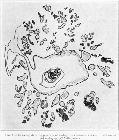
Fig. 1. Drawing showing position of embryo in chorionic vesicle. Section 95 (of embryo). X 15 diameters.
The chorionic vesicle appeared as an elongated rounded body, of a cream color and a very delicate texture. It measured 15 by 9 by 8 mm. Extending from end to end of the vesicle, a flat fold with a slit in it was visible. It was covered with villi everywhere except along the fold. For fear of ruining a valuable embryo no attempt was made to open the sac. The whole vesicle was run through the graded alcohols, cleared in chloroform, and cinbcddcd in parafiin. It was cut in sections 8 microns thick and stained with alum hemotoxylin and eosin. An idea of the plane of sectioning can be obtained from text-figure 1.
The age of the specimen is uncertain from the data obtained. As determined from a model of the embryo, its greatest length is about 2.4 mm. This measurement and those of the chorionic vesicle make it correspond very closely to an embryo of 28 days, as estimated by Mall'^, p. 199). If such is its age the specimen falls into that group of embryos w^hich continue to develop in the uterus during a menstrual flow.
Comparison with descriptions of other embryos shows that my specimen is near the age of Robert Meyer's embryo No. Meyer 300, described by Thompson"; it resembles also Janosik's specime^" and His's embryo "Lg."*. In most respects, however, it is younger than the above-named specimens and also younger than the embryos described by Mall^', Gage'^, Bremer^, and Tandler^^ It is apparently older than the twin specimens of 17-19 segments described by Watt**.
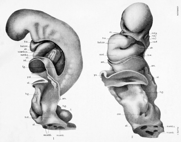
A glance at the figure (plate 1, fig. 1) of the embiyo is sufficient to show that there is a marked ventral bend in its back, such as has been found in many specimens of comparable age. Since this ventral flexure is not invariably present, and varies in degree, it is regarded by Keibel as essentially abnormal. In my embryo it occurs without rupturing the underljdng structures; if an abnormality, it is the only one which seems to be present.
I am indebted to my former students, Messrs. . L. Brosius, L. B. Hohman, H. L. Houchins, L. II. Rutledge, Florien Vaughn, and T. F. Wheeldon, for certain reconstructions which have aided me greatly in my study of the embryo. To these men and also to Mr. G. T. Kline, who has made many of the illustrations, I wish to express my sincere thanks. I wish also to express my gratitude to Professor Frederic T. Lewis, who has read my manuscript and offered many valuable criticisms.
External Form
The external form of the embryo has been studied from a wax reconstruction which was made at a magnification of 120 diameters. As already stated, the embryo was cut up without being previously drawn. It was necessary, therefore, to use the walls of the chorionic sac as guide-lines in making the model.
As seen from the left side (plate 1, fig. 1), the embryo is roughly the shape of a reversed S. Its back presents two well-developed curves. The upper of these is convex dorsally; it is large and rounded. The lower curve is convex ventrally, since the caudal portion of the embryo is bent sharply backward at about right angles to its longitudinal axis. Not only is the caudal end of the embryo bent backward upon itself, but at this joint of bending it is twisted through an angle of 90° to the right. Thus the dorsal wall of this portion of the embryo is turned to the right and the ventral wall to the left; also, the caudal end of the embryo is directed to the left, so that its tip lies to the left of the axis of the body of the embryo.
The head of the embryo is large and rounded. When viewed from in front (plate 1, fig. 2), it appears somewhat egg-shaped. Toward its middle, on either side, are distinct outward bulgings, beneath which lie the optic vesicles. The mouth is a well-defined opening, limited on each side and below by the large mandibular arches.
Behind each mandibular arch there are three distinct gill-clefts. The first two of these are long and narrow and are directed obliquely to the longitudinal axis of the body, but the second bends dorsalward in its upper portion at almost right angles; the third is more shallow and rounded. The gill-clefts are bounded by four distinct arches. The first of these, the mandibular, is large and rounded, the second is similarly shaped but smaller, the third and fourth appear merely as rounded eminences.
The pericardium and lieart lie just ventral to the arches. They form a large bulging which is more prominent on the right than on the left.
Beginning some little distance caudal to the last arch and placed at regular intervals throughout the remainder of the embryo, the mesodermic somites can be seen bulging through the skin. About 15 of these are perceptible from the surface, 10 in front and 5 behind the sharp flexure of the back.
The amnion is reflected from the embryo at the lower end of the pericardial cavity. The line of reflection here is curving and follows the lower curvature of the pericardial wall. At the side of the embryo, about halfway between the dorsal and ventral mid-lines, the line of reflection turns caudally, passes along the sides of the yolk-stalk, along the sides of the embryo, and finally on the body-stalk. Thus all the head, pericardial wall, and most of the caudal extremity and the dorsal portion of the remainder of the body of the embryo are within the amnionic sac.
Ventral to the line of reflection of the amnion is the yolk-sac. This is a large, irregular vesicle, broken through in places and flattened laterally. The yolkstalk, which proceeds from the embryo just beneath the pericardial sac, is also flattened, but from above downward.
The outer surface of the hind gut, i. e., its mesothelial surface, is distinctly seen turning backward to follow the curvature of the caudal extremity At its bend there are two projections of the body-cavity, which likewise pass backward into the tail.
The body-stalk is attached to the embryo near its lower end. ventral to the caudal extremity. It turns sharply backward. The amnion is reflected from its upper surface for a short distance beyond the embryo. As a whole the body-stalk is short and broad, gradually becoming broader as the chorion is neared.
No indications of either fore or hind limb-buds can be found on the body of the embryo.
In comparing this embryo with Janosik's, it is seen that the back of the latter presents a more even curvature, which extends to the tail of the embryo. The angle formed at the top of the head (the midbrain bend) is almost identical in both specimens. The head of Janosik's embryo, however, is pointed, while in my specimen it is more rounded. In marked contrast to both of these specimens is the Robert Meyer embryo No. 300, as modeled by Thompson. Here the head bend is very gradual, and the head itself much narrower and more pointed than in either Janosik's specimen or mine. Although somewhat similar in appearance to His's'" embryos "Lg." "Sch," and "BB," my specimen differs from them in regard to the position of the ventral flexure of the back. In His's specimens the much-discussod bend in the back is always placed opposite the attachment of the yolk sac and stalk. In my specimen, however, the bend is placed further caudally, and the portion of the body which is bent backward is relatively shorter. The twin specimens which Watt" describes show definite ventral curvatures of the back, but these also are placed relatively higher up and are not as sharp as the bend in my specimen.
Integument
In general, it may be said that the integument of the embryo is made up of one or two layers of ectodermal cells. The thickness of this layer and the shape and size of its cells, however, vary considerably in different regions of the body.
Over the sides of the head the ectoderm is thin, being composed for the most
part of two layers of flattened cells with rounded nuclei. On the dorsum and
front of the head the epithelium is still thinner, there being but one layer of
flattened cells. Over the optic vesicles, where the lens placodes will later develop,
there is as yet no indication of thickening, but in the region of the gill-arches the
epithelium is considerable thicker, its cells being either cubical or columnar in shape.
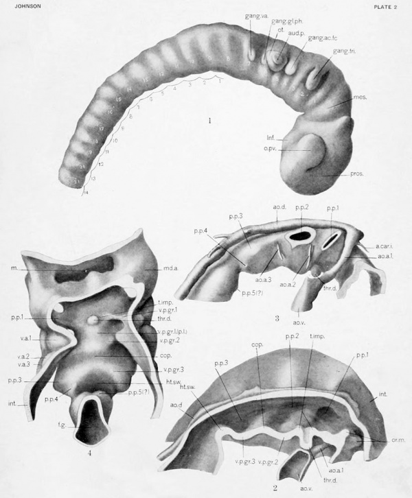
In the region of the hindbrain there is seen from the surface a minute aperture (plate 2, fig. 1). Here the integument dips in and expands to form a sac,
the auditory vesicle. This is flattened laterally, and approximateh' triangular
in external view. It is closely applied to the brain, overlying the fifth and a part
of the sixth neuromeres. With the exception of its form it is quite closely in
accord with the more spherical vesicle of Thompson's embryo. The walls of the
auditory vesicle are much thicker than the overlying ectoderm, and exhibit two
or three layers of rounded or oval nuclei. Mitotic figures in the auditory vesicle
are numerous.
The integument in the region of the mouth shows no especial thickenings.
A few clusters of cells (the remains of the oral plate) are attached to it; anteriorly,
one such cluster is found at about the level of the cephalic end of the notochord;
other clusters are found on the sides and ventral wall of the oral cavity. The
epithelium of the roof of the mouth is placed in close apposition to the floor of the
fore])rain, being separated from it by a few strands of mescnchyma only. There
is no doubt that this portion of the oral integument is destined to become the
anterior lobe of the hypophysis, but as yet there is no definite differentiation of
this organ.
The integument of the body-wall of the embryo overlying the pericardial cavity is very thin, suggesting a stretching-out of the epithelium. It is also thin
over the dorsum of the trunk and over the mesodermic somites. The integument
which overlies tlie body-wall in the region of the umbilical vein is somewhat
thicker and its nuclei are more closely packed together. It gradually thins out
again as it is reflected to form the amnion.
The Nervous System
The nervous system is represented Ijy the brain, with its two optic vesicles and the beginnings of the trigeminal, acustico-facial, glosso-pharyngeal, and vagus nerves, and the medullary tube with its ganglionic crest. The dorsal wall of the nervous system is placed just beneath the integument of the mid-dorsal line and conforms to all its curvatures. The cavity of the tube is entirely closed off from the outside, disregarding a longitudinal slit in the mid-dorsal line, which is clearly artificial, and in this respect differs from that of Janosik's embryo, which showed a small anterior neuropore.
The Brain
The three primary vesicles of the brain are easily recognizable in plate 2, figure 1. The prosencephalon is large and bulbous, and is marked off from the mesencephalon by a deep groove. Broad in front and in the region of the optic vesicles, it gradually becomes narrower behind. Its cephalic end is rounded and lies in close contact with the ectoderm; an actual fusion is at one place apparent, marking probably the position of the closed anterior neuropore. There is no indication of a hemispheral division. The optic vesicles are attached to this portion of the brain slightly ventral and anterior to its middle; then extend outward, backward, and sUghtly dorsalward. There is as yet no definite indication of a division of the prosencephalon into diencephalon and telencephalon. A slight rounded protuberance of the ventral wall behind the points of attachment of the optic vesicles probably marks the beginning of the infundibulum.
The mesencephalon is a small wedge-shaped portion of the brain-tube. Of the grooves which mark it off from the prosencephalon in front and the rhombencephalon behind, the anterior is deeper; the posterior groove is faint dorsally, but ventrally it ends in a deep notch. The mesencephalon is much narrower from side to side than the prosencephalon. Its antero-posterior dimension is only a trifle greater than those of the neuromeres of the rhombencephalon immediately behind it. In marked contrast to this is the much longer and larger mesencephalon of the Robert Meyer embryo, as modeled by Thompson'^ Somewhat similar to it, however, is that of Ingalls's embryo.
The rhombencephalon is elongated and flattened laterally. As seen from the side it is slightly curving, its dorsal wall being convex.
Medullary Tube
The spinal part of the medullary tube extends from the rhombencephalon to the tip of the tail, gradually tapering from above downward. It is ovoid in section, the lateral walls being thicker than the dorsal and ventral walls. The dorsal wall is thinnest and lies almost in contact with the covering ectoderm.
Neuromeres
Both the rhombencephalon and spinal portion of the medullary tube are marked off by transverse grooves into a series of segments, the so-called "neuromeres." These begin at the cephalic end of the rhombencephalon and continue downward through the medullary tube. The first six neuromeres are narrow.
Lying next to the second neuromere is the ganglion of the trigeminal nerve; next to the fourth neuromere is the ganglion of the acustico-facial. The auditory vesicle lies opposite the fifth neuromere and partly overlaps the sixth; the latter is in process of giving off the cells of the glosso-pharyngeal ganglion. Beginning with the seventh, the remaining neuromeres are longer, being equal in length to the body segments. They do not lie within the segments themselves, however, but are arranged intersegmentally, the crest of each neuromere being placed opposite an intersegmental cleft. This arrangement begins with the first body segment and continues throughout the embryo. Above the first segment are 8^ neuromeres.
The number of neuromeres belonging to the rhombencephalon can not lie ascertained from this specimen alone. To determine this point, the author undertook a separate study of neuromeres based upon young human, pig, sheep, and cat embryos. Although this study is yet incomplete, it is evident that the last rhombic neuromeres stand out more clearly in certain specimens than in others; in some their presence is extremely doubtful. Just what this may be attributed to I am not able to state; it may be due to the stage of the specimen examined, or their presence on the one hand or absence on the other may be regarded as artificial. In those specimens which show distinctly a complete series of neuromeres, I have found that the first cervical ganglion is constantly related to the tenth neuromere. It appears evident, therefore, that the first 9 neuromeres belong to the rhombencephalon. It is to be noted that the first pair of somites in the embryo under discussion begins approximately opposite the crest of the eighth neuromere.
Of the previous descrijition of neuromeres in young human embryos, those which appear to accord most closely with my own are the ones of Clage and Watt**. In a description of an embryo of 28-29 pairs of somites, ^Irs. CJage finds 9 folds or neuromeres in the rhombencephalon; of these the second is associated with the trigeminal nerve; the fourth with the auditory and facial nerves; the fifth is opposite the auditory vesicle; the sixth is in relation to the glosso-pharyngeal nerve; the seventh to the vagus; and the eighth and ninth to the accessory nerve. In regard to the spinal cord she states:
- "Beyond the clearly fonned folds, above discussed, there occur several others, each corresponding with an enlarged part of the ganglionic cord. As this cord has no further indication of dorsal nerve roots, the exact relations ran not be determined. Moreover, the following total folds in the niyel (spinal cord) are not strongly marked, and in other spefimens it is only in favorable sections that they can be seen at all." (pii. 435-430.)
Watt", in describing twin human embryos of 17-19 pairs of somites, similarly shows 9 neuromeres in the rhombencephalon. The tenth neuromere, he states, is opposite the first cervical ganglion. The results I have obtained with other mammalian embryos confirm this observation. Watt also shows spinal nenromeres extending along the medullary tulx^ as far as the eleventli spinal ganglion.
In 1892, Minot" reviewed the earlier literature on this subject and made the general statement that "the entire medullary tube undergoes a segmentation by a series of alternating slight enlargements and constrictions." He adds:
- "They appear first in the hind-brain and cervical region, and from there they appear progressively toward the fore-brain and the tail .... The medullary tube becomes slightly constricted between each pair of segments and shghtly enlarged opposite each intersegmental space. Each intersegmental dilation is a neuromere .... Each neuromere produces a pair of nerves, but when the first trace of roots appears, they are seen to spring from the constriction between the neuromeres, but later from the neuromere."
Minot believes, therefore, that the so-called neuromeres of early stages represent, not true neuromeres, but the caudal and cephalic lialves of two adjacent neuromeres.
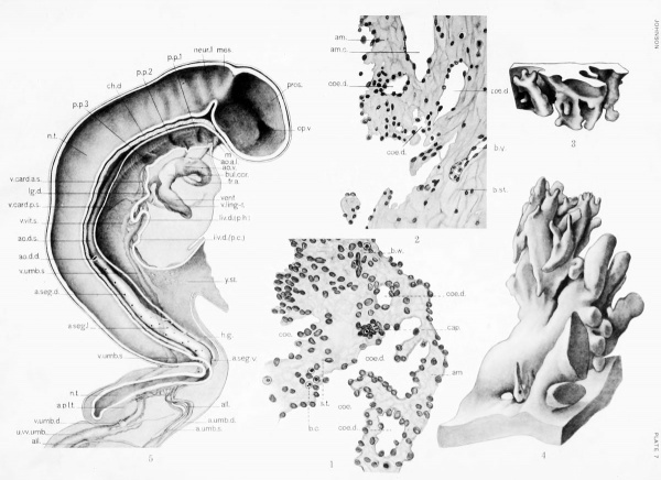
An internal view of the brain is shown in plate 7, figure 5. The cavity of the prosencephalon is large and deep. Towards its anterior end the cavity of the optic vesicle is seen extending outward and backward. Posteriorly this is marked off from the forebrain cavity by a sharp indented ridge. The slight eminence of the ventral wall which will probably give rise to the infundibulum is again seen, being placed at the anterior end of the notochord. The walls of the rhombencephalon, when viewed from within, show the negative impression of the neuromeres, the "rhombic grooves" of Streeter".
The question of neuromeres in mammaUan embryos is still an open one. Those of the spinal cord are undoubtedly related to the metameres, i. e., representing true or parts of true morjjhological units of the medullary tube. Similar evidence concerning the "neuromeres" of the rhombencephalon is lacking. Although the arrangement of cerebral nerves is not contradictory to this view, ?'. e., each neuromere, with the exception of the first, receiving afferent fibers and sending out efferent fibers (Johnson)'*, the muscles which the efferent nerves supply, the source of these muscles, and the "neuromeres," from which the efferent nerves spring, have not yet been demonstrated to coincide. In fact, the evidence, so far as gathered, contradicts this arrangement. Streeter"*^ believes that the rhombic folds are not related to the metameric system, but is inclined to the view that "they may be fitted in with and form a part of the branchioraeric system."
He adds:
"The one discordant feature is groove d (5th neuromere), which has no corresponding branchial arch."
NeaP, after extended observations and studies of head segmentation, in embryos of lower vertebrates, concludes that neuromeres offer no criterion for the determination of segmentation of the vertebrate head.
Cerebral Nerves
As in the Janosik and Thompson embryos, the ganglia of the trigeminal and acustico-facial nerves are clearly present. Of the two, the trigeminal arises a little higher dorsally than the acustico-facial. Each is made up of a cluster of nuclei which are more compact and deeply staining than the mesenchymal cells which surround them. They are connected with the wall of the brain by strands of cells; definite fibers are apparently just beginning to form. Distally, ganglion cells and fibers can be traced for some little distance, the trigeminal nerve passing toward the maxillary and mandibular arches and the acustico-facial into the dorsal portion of the second arch.
In addition to the trigeminal and acustico-facial nerves, there is found just behind the auditory vesicle and near the brain-w'all a small cluster of undifferentiated cells. These are found in the region of the sixth neuromere and very probably represent the ganglion of the glosso-pharyngeal nerve. Opposite the seventh neuromere is another similar group of cells, the probable beginning of the vagus nerve.
Ganglionic Crest
Indistinctly' connected to the vagus ganglion and extending down beyond tlic ventral bend in the back of the embryo, a ganglionic crest is discernible. It is not distinct, however, for its cells so closely resemble those of other adjacent tissues that they can not always be identified with certainty. It is largely due to the jiosition of its cells, i. e., between the medullary tube and the somites, and to the arrangement of its nuclei, that the jiresence of the ganglionic crest can be detected.
Streeter" has described the neural crest of a 4 mm. embryo as follows:
"This structure (the ganglionic crest) can be seen in the 4 nim. embryo as a flattened cellular band which extends caudahvard from the auditory vesicle along the lateral wall of the neural tube to its extreme tip .... That part of the crest whicli corresponds to the spinal cord is characterized at this time by segmental incisures along its ventral border. The dorsal border of the crest remains intact until the appearance of the dorsal rootlets, in the meantime constituting a cellular bridge coimecting the more ventral ganglionic clumps."
In my specimen the ganglionic crest likewise forms a cellular band. Segmental incisures along the ventral border are indicated in certain regions. The crest, however, is apparently not so far developed as shown by Streeter. Caudally its development is not so far advainced as toward the head. So indistinct and uncertain are its outlines, as seen in sections, that I have made no attempt to rejiresent it graj)hically.
Lenhossek^'^ has show'u the ganglionic crest in a formative stage in a human embryo of 13 segments. Its cells are described as arising from the roof of the medullary tube. Such pictures as Lenhossek has shown I have been able to find in my specimen only in the caudal i)ortion of the body, where the roof of the medullary tube is relatively thick; there, as seen in cross-sectif)n, the develoi)ing ganglionic crest ajjjjcars as a cap lying on the roof of th(> medullary tube; it is coniposefl of closely i)acked cells with luulci of about the same size as those of thf remainder of the tube.
Digestive System
Mouth
The mouth has been partly described in connection with the integument. It is directly continuous with the pharynx, the line of former separation being represented above by a few scattered clusters of cells, and below by a thin ridge of epithelium, remnants of the oral membrane. The oral cavitj' is broad transversely but narrow dorso-ventrally; there is no indication of the anterior lobe of the hypophysis. The scattered cells which Janosik'" has designated as the hypo))hysis are undoubtedly remnants of the oral membrane.
I shall here mention His's'* description of his embryo "Lg" (2.1.5 mm.), in which he recognizes both Rathke's and Seessel's pockets, wliile the oral membrane is still intact. Concerning these he says:
"Of the two peaked recesses between which it (oral membrane) passes, the anterior becomes Rathke's pocket, while posterior becomes Seessel's pouch."
In an embryo of 2.6 mm. in Keibel and Elze's NormentafeP', in which the pharyngeal membrane is still present, the hypophysis is "just indicated." The Robert INIej'er embryo of 23 segments, according to Thompson, shows no hypophysis, although the Normentai'el states its beginning is "doubtful."
Foregut
Pharynx
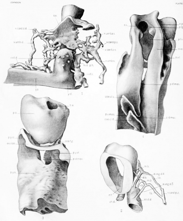
The cavity of the pharynx is broader and deeper than that of the mouth. In the median plane its dorsal wall lies ventral to the notochord and follows closely the curvatures of that structure, being fused vrith it posteriorly (text-fig. 7). Ventrally the floor of the pharynx is more irregular. It possesses, toward its anterior end, a short, rounded diverticulum, the beginning of the thyroid gland plate 2, figs. 2, 3, 4). This is in close relation ventrally to the ventral aorta, with which it lies in contact. Thompson describes a thyroid gland which is apparently in a similar stage of development, but Janosik and His (in his embryo Lg), fail to show this organ.
Behind the thyroid diverticulum the ventral wall of the pharynx shows two shallow depressions which cross the midline (plate 2, fig. 4). These are the cut sections of the transverse grooves (the "ventral pharyngeal grooves" of Grosser) which extend from side to side and connect the pouches of one side with those of the other. As described by Grosser*, I find that the ventral groove of the first pouch before reaching the midline divides into two limbs which surround a median elevation. This he identifies as the "tuberculum impar." It is to be noted that the thyro-glossal duct proceeds from the summit of this elevation. This relation is noted by Grosser, who describes it as follows :
- "The opening of the thyroglossal duct is situated at first upon the summit of the tubercle, but later it becomes shifted into the furrow bounding the tubercle posteriorly or, according to Ingalls, in an embryo of 4.9 mm., into the region of the second arch, immediately aboral to the tuberculum impar.' "
In a study of two embryos of 3 mm., Hammar17 reports the tuberculum impar present in only one.
Between the ventral pharyngeal grooves of the second and third pouches is a second medial elevation in the form of a transverse ridge. From its position I believe it to be the so-called "copula" which, according to Ilis and others, helps to form the tongue.
A third prominent elevation is found behind the ventral pharyngeal groove of the third pouch. It marks the point where the pharynx turns sharply caudalward. This probably is the area which Grosser had denoted the "cardiac swelling" and which he states goes into the formation of the larynx.
|
|
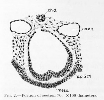
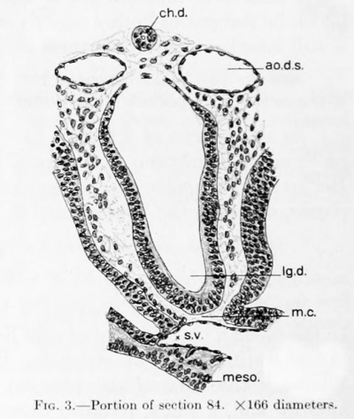
| Fig. 2. Portion of section 70. X166 diameters. | Fig. 3. Portion of section 84. X166 diameters. |
The lateral borders or wall of the pharvnx extend outward into three distinct pharyngeal pouches. Of these, only the first and second reach the ectoderm. The first pair of pouches is situated only a short distance behind the oral membrane. They are flattened from before backward, the left being somewhat broader than the right. In studying the first pouch of young embryos, (Jrosser'* describes an invagination of its epithelium into the pharyngeal cavity as follows:
- "In the region of the first pouch tlicro projects ventrally or caudally from the closing membrane into the pharyngeal lumen an irregularly knotted process filled with mesoderm .... It disappears quite early and may perhaps be interpreted as a rudimentary internal gill."
I have looked carefully in the region designated by Grosser for the structure which he describes, but have been unable to hnd any dehnite indication of it. A slight irregularity of the epithelium, however (more distinct on the left side than on the right), corresponds with it in position.
The second pouch is somewhat larger than the first and is flattened dorsoventrally. A distinct ventral diverticidum can be .seen. The third pouch is more rounded in form and ends bluntly in the mesenchyme. falling somewhat short of reaching the octodoriii. A fourth pouch is seen extending from the ventro-lateral surface of the pharynx. It arises to a certain extent in common with the third, and shares with it the third ventral pharyngeal groove. It is slightly pointed and is directed outward, backward, and downward.
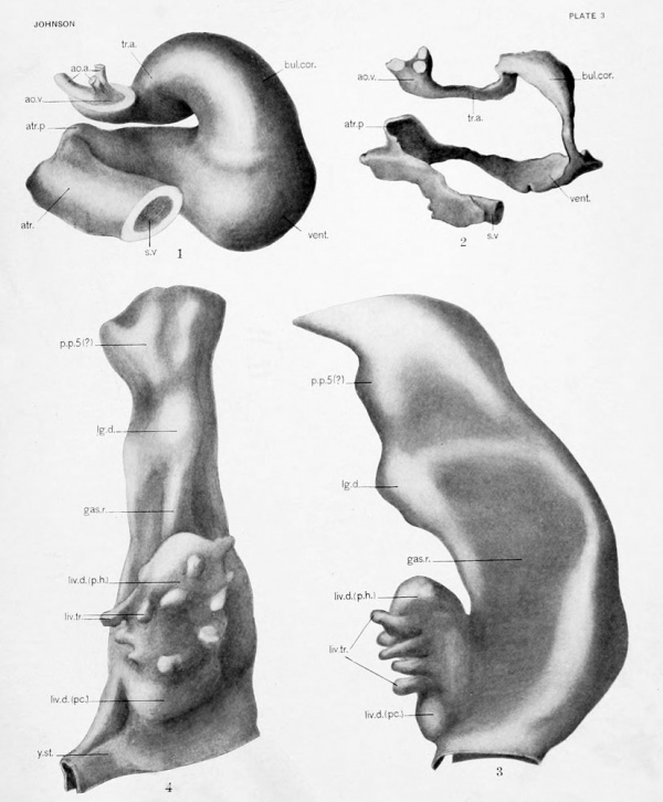
The remaining portion of the foregut, that is, that part between the fourth pharyngeal pouch and the yolk-stalk, is shown in plate 3, figures 3 and 4. It represents, above, a definite swelling which is apparent when seen in either front or side view. Looked at from in front, the swelling appears double, a median longitudinal groove separating two rather elongated protuberances. A crosssection of this region is shown in text-figure 2. A short distance below this swelling there is another which, when viewed from the side, is seen to be rather pointed. It is just dorsal to the sinus venosus (text-fig. 3). Still farther caudally is seen the hepatic diverticulum (text-fig. 5).
|
|
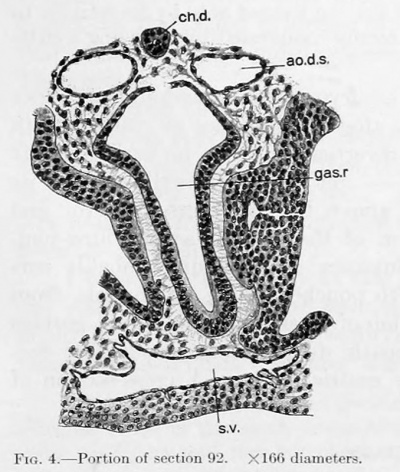
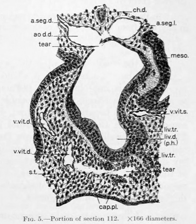
| Fig. 4. Portion of section 92. X166 diameters. | Fig. 5. Portion of section 112. X166 diameters. |
Pulmonary Diverticulum
The significance of the two upper swellings I am unable to determine definitely from this specimen alone. Thompson" describes a somewhat similar condition in the Robert Meyer embryo of 23 segments. The upper swelling he interpreted as a bilateral pair of lung-buds; the lower as the beginning of the stomach. Grosser'^, however, from a study of this region of the same embryo, concluded that Thompson's bifid swelling represents probably a fifth pair of branchial pouches, and that the lower .sweUing is the lung-bud.
Regarding Thompson's description he states :
- "Thompson (1907) asserts that in this embryo the lungs have a paired origin, but he does not figure it, and this statement has been transferred to the Xormentafel. But he is clearly in error as to the place where the huigs develop, as shown by liis own description and a figure which he pubhshed later (1908). In 1908 he wrongly identified what is actually the king-bud as the stomach, and 1907 he placed the lung-bud in the region of the diverticulum, which I have identified as a questionable fifth pharyngeal pouch. In fact, the embryo has as yet no indication of the stomach. Moreover, liis model was probably made on too small a scale."
The longitudinal groove which Grosser'^ describes extending from the region of the last jjouch down to the lung-bud is not present in my specimen.
According to Lewis^, the Bremer embryo pos.sesses a definite pyriform lungbud, which is directed ventrally and caudally. The Broman embryo of 4.25 mm., as figured bj' Grosser'*, shows a similar lung-bud.
Gastric Region
For the following unpublished statement concerning the gastric region of the Bremer embryo I am indebted to Professor Lewis :
- "In the Bremer embryo a gastric region may be referred to, but it is not marked off in any way from the esophagus or duodenum. It may be located only by its relation to the liver and body cavities. It is a flattened or laterally compressed tube, having a cleftlike lumen."
From the condition found in the Bremer embryo, which is undoubtedly older than mine (having limb-buds), it would seem that the presence of a stomach in my specimen is improbable. A pulmonary diverticulum is to be expected. It seems to me, therefore, more reasonable to interpret the lower swelling, from its relation to the hepatic diverticulum, lying above the transverse septum and
consequently in the jileuro-pericardial portion of the coelom, as the lung-bud. The divided swelling situated above the j^ulmonarj' diverticulum probably corresponds to Grosser's doubtfully identified fifth pouches, but I am unable, from my specimen, to confirm Grosser's interpretation of its significance. That portion of the foregut between the lung-bud and hepatic diverticulum presumably corresponds to what Lewis^ has designated the gastric region. A cross-section of this region is shown in text-figure 4.
Hepatic Diverticulum
A short distance below the gastric region the gut widens out considerably, forming a third diverticulum, the liver (text-fig. 5, and plate 3, figs. 3 and 4). It is directed ventrally and orally. It presents at about the junction of its lower and middle thirds a broad, shallow, transverse groove which divides the diverticulum into two iiortions, both entirely embedded in the septum transversum. Extending ventrally and laterally fnnii llic \i\)\)rv portion are a number of buds, the beginnings of hepatic trabeculae; no such buds arc found in connection with the lower part. The buds are comi)osed of proliferating epithelial cells which, as indefinite cords, have invaded the mesenchyma of the septum transversum. Most of them are indistinct and their extent is doubtful, for their cells closely resemble those of the mesenchyma and there is no definite line of separation between the two, such as a basement membrane. They are readily overlooked, and it is only with an oil-immersion lens that they an; made out witii any degree of certainty.
Felix" notes the same difficulty in tracing hepatic trabecular in a slightly older embryo. Careful study shows that the nuclei of the entodermal cells are slightly larger than those of the mesenchyma, a point that is helpful in determining which cells belong to the trabecular.
The Bremer embryo (4 mm.) shows a somewhat more differentiated stage in the development of hepatic trabeculse. Here they form anastomosing cords (Lewis)^*. As shown by Ingalls" in a 4.9 mm. embryo, the trabeculse are very extensive and form a large mass of anastomosing cords. Thompson^, however, found no evidence of hepatic trabeculse. In a later note" he states that "the transverse septum is seen before the cells of the liver bud have invaded the vessels which lie in it." He shows in sections, however, a transverse septum which, like the one in my embryo, is ciuite thick, and he apparently considers that all its cells (excluding endothelial and blood cells) are mesenchymal. Judging from the definiteness and size of the hepatic trabeculae of the Bremer^ embryo, which is evidently only a trifle older than Thompson's or my own, one would naturally expect to find some evidence of the trabeculse in the latter two. I believe that, owing to their indistinctness, it is possible that they were overlooked by Thompson.
Regarding the development of hepatic trabeculse, I believe it safe to draw the following conclusions: that they arise as indefinitelj^ outhned buds of proliferating entodermal cells from the upper portion of the hepatic diverticulum; that while they grow in the mesenchyma and anastomose with one another, their cells undergo further differentiation and thej' become more distinctly differentiated from the mesenchyma.
I must mention briefly at this i)oint the relation of the hepatic trabeculse to the veins of the transverse septum. Janosik'" has noted that the early hepatic diverticulum in the human embryo is not related to the vitelline veins in the same way as in birds. Bremer^ states:
"The liver cords are found growing into the mesenchjTna, at a level ventral to the vitelline veins; in this same mesenchyma, however, we find the branches of the vitelline veins ramifying and forming plexuses, and in certain places these plexuses come into intimate relation with the liver cords."
I find with Janosik and Bremer that the hepatic diverticulum and trabeculae are not in close relation to the vitelline veins. These lie dorsally and laterally to the diverticulum. Somewhat ventrally and anteriorly is found the sinus venosus. In the region of the hepatic trabecular can here and there be made out minute spaces which contain one or two red blood-corpuscles, apparently blood-vessels; but in my specimen I have been unable to make out a definite plexus as found by Bremer.
The lower knob-like portion of the hepatic diverticulum presents no special feature other than a very thick ventral wall. Brachet', in a careful study of the development of the liver in several different vertebrates, shows that in the rabbit the hepatic diverticulum is an elongated outpocketing of the foregut.
extcndinji from tlio ri'sioii of the sinus venosus to the yolk-stalk, and divisil^le into cranial and caiulal portions. The upi)('r of these he states goes into the formation of the liver proper and the hepatic duct; the lower into the formation of the gall-bladder and the cystic duct. The two portions are designated by Maurer" the "pars hepatica" and "pars cystica" respectively. The twin embryos which Watt described are apparently too young to show these divisions of the liver— in fact, the liver forms merely a slight swelling on the ventral wall of the foregut where the latter joins the yolk-stalk. It is called by Watt the "liver bay." Thompson, however, recognizes the hepatic and cystic i)ortions of the liver diverticulum in the Robert Meyer embryo No 300. Somewhat similar divisions are described by Ingalls in his embryo of 4.9 mm., but his specimen is considerably older than the above-mentioned ones. Evidence of a division into two portions is apparently altogether lacking in the Bremer embryo of 4 mm., the liver diverticulum of which has been modeled by Bremer and more recently by Lewis.
Yolk Stalk and Sca
Just below the he])atic diverticulum the gut becomes narrow, but a little more caudally it again gradually broadens. This broadening leads out into the cavity of the yolk stalk and sac. The yolk-stalk is short, being in fact merely the constriction between the yolk-sac and the gut. It is flattened antero-posteriorly, but is broad transversely (plate 1, fig. 2). It measures roughly 0.5 mm. from side to side and 0.16 mm. antero-posteriorly.
The yolk-sac is a flattened vesicle, rather irregular in form, and with a number of folds of various shapes and sizes on its surface. It fills up j^ractically the entire apaee between the embryo and the wall of the chorion, and extends into the artificially made fold of the chorionic wall as described above. Its dimensions are roughly as follows: length, measured parallel to long axis of embryo, 3.3 mm.; width, measured parallel to dorso-ventral axis of embryo, 2.7 mm.; thickness, measured transverse to embryo, 1.1 mm. Its histological structure will be considered later.
Hind Gut and Cloaca
Caudal to the place at which the yolk-stalk passes out is the beginning of the hind-gut. It has a funnel-shaped ojjcning which taj^ers as it passes toward the tail into a small rounded tu])ule. It is surrounded by loose mesenchyma, the wliole being attached to the df)rsal body-wall by a short, thick mesentery. The hind-gut occui)ies a position slightly to the left of the median plane of the embryo. It bends backward with the body of the embryo at the ventral bend in the back. It passes without sharp demarcation into the cloaca (text-figs. 8 and 9). The cloaca is the cejjhalic portion of the spindle-shaped termination of the hind-gut. Its cej)halic limit is not definitely indicated, but its caudal extent is marked by the cloacal membrane.
Allantoic Duct
The allantoic duct is a very long, slender, hollow tube which proceeds from the cephalic end of the cloaca and extends into the body-stalk. At its origin it is funnel-shaped, and its lumen is distinct. It tapers rapidly as it enters the bodystalk. For a few sections it becomes almost lost from view, owing to the indistinctness of cell boundaries and scattered nuclei. Although continuity of the allantoic cells can be made out, its lumen is lost. A few sections farther distally the allantoic duct again becomes distinct and a trifle larger. Lying between the two umbilical arteries, it follows the ventral bending of these vessels. Still lower down the arteries fuse and then split apart again, thus forming an arterial fork. The small allantoic duct passes in front of the fused part, and then, turning dorsally, passes through the above-described fork. Crossing the umbilical stalk obliquely, it terminates in a small bulb, the allantoic vesicle, which is situated close to the fused umbilical veins.
CLOACAL MEMBRANE.
A short distance from the end of the gut is a very slight outward bulging of the ventral wall. This portion of the cloacal wall is in contact with the ectoderm of the proctodeal invagination, and together these layers of epithelium form the cloacal membrane (text-figs. 8 and 9). The entodermal portion is slightly thicker than the ectodermic.
CAUDAL INTESTINE.
The portion of the gut beyond the cloacal membrane ends bluntly in the extreme end of the tail, separated from the ectoderm by only a small amount of mesenchymal tissue. This portion of the gut represents the post-anal or caudal intestine (text-figs. 8 and 9).
Histological Structure of the Digestive Tract
Histologically considered, the digestive tract maj' be described as an epithelial tube surrounded by mesenchj^ma. Only the former shows signs of differentiation as yet, the mesenchjTna being everywhere of the same character. The epitheUum takes on widely different appearances in different regions. In general it may be said that throughout the whole of the digestive tube, with the exception of the lower end, the dorsal wall is much thinner than the ventral. The former is made up of a single laj'er of cubical or flattened cells. On either side of the mid-dorsal line the epithelium gradually becomes thicker and the nuclei more crowded. The side-walls and floor of the pharynx show from two to three layers of nuclei. At the places where the entodermal epithelium of the pharyngeal pouches comes in contact with the ectoderm, it is thin and fused to the ectoderm. The membrane closing the second gill-cleft on the left side has broken through to the outside; this is undoubtedly a mechanical tear. The wall of the thyroid diverticulum is not different iiom that of the floor of the pharynx, being composed of an epithelium of two to three cell-layers. The epithelium of the ventral wall of the remainder of the fore-gut is thicker, the change from the thin dorsal wall to the thick ventral wall taking place gradually on the sides of the tube. The pulmonary diverticulum is two to three cell-layers thick, the hepatic diverticulum three to four.
In the region of the yolk-stalk the epithelium is made up of one layer of cubical or somewhat flattened cells. In the yolk-sac the epithelium is not everywhere the same. In some places the cells are large and cubical; in other places flattened and less distinct. Often is it impossible to determine whether or not an epithelium is present, for in such places the epithelial cells can not be distinguished from those of the mesenchyma. The mesenchyma surrounding the yolk-sac also varies in thickness. It contains numerous blood-vessels and is covered by the mesothelium of the body-cavity.
The hind-gut, down as far as the cloaca, is composed of but a single layer of cuboidal cells, which is of eoiual thickness all around. In the cloaca and caudal intestine the epithehum of the side and ventral walls is thickened, and is coml)osed of two to three layers of cells. The entodermal epithelium of the cloacal membrane shows no distinguishing characteristics. It abuts against the ectodermal epithelium, but both layers can be made out distinctly.
Somites
In all, 24 pairs of somites are present; they extend from the lower end of the hindbrain to a httle beyond the point where the allantoic duct passes out from the cloaca. The first somite is found in the region of the eighth and ninth rhombic neuromeres. According to studies on a slightly older human embryo and on young embryos of the pig, sheep, and cat, I have found that this position is normally occupied by the second somite. Watt^* likewise show^s the second somite in this position, the first somite being in relation to the seventh and eighth neuromeres. I have looked repeatedly in my embryo, however, for evidence of another sormte in front of the one which I have designated as the first, but have been unable to find any definite indication of such.
As in other young embryos which have been described, the different pairs of somites are found in different stages of development, those nearer the head end always being more advanced than those behind them. In the caudal end of the embryo are found somites in the process of formation; in the head they are already partially broken up. Following Ingalls's plan, I shall begin my description with the somites of the tail and proceed forwards.
The caudal end of the vertebral plates of mesoderm till up entirely the tail of the embryo around the neural tube, notochord, and tail gut. In the region of the cloacal membrane they api)ear as solid masses or cords of mesoderm with closely packed cells. At their cephalic ends a small cavity is apparent. Another pair of somites, the twenty-fifth, are i)artially formed by an incomplete transverse furrow. Numerous mitotic figures are present in the vertebral plates.
The twenty-fourth somite (second lumbar) is in a very early stage of development. It is somewhat cubical in shape and in its cenler is a distinct cavity, the myocoele. The walls of the somite, whidi iiiny be dtscribed as dorsal, ventral, medial, and lateral, are all of about equal thickness. They are epithelial in character and contain one to two layers of cells. In the myocoele are found a few scattered stellate cells not unhke mesenchymal cells; later these will enter into the formation of the sclerotome. Mitotic figures are numerous among the cells of the walls and the myococle, but those of the walls are always at the upper ends of the cells, i. e., the ends bordering on the myoccele. This somite corresponds quite closely with the first coccygeal somite of the Ingalls embryo, except that it probably has fewer cells in its cavity.
The nineteenth somite (ninth thoracic) shows a somewhat more advanced condition. The myoccele in its lower portion is entirely filled with cells. The ventral half of the medial and all of the ventral wall arc breaking up. The cells of these walls, together with those on the inside of the myoccele, form the sclerotome. These cells have pushed out slightly toward the chorda dorsalis, forming the notochordal process. The somite corresponds with the sacral somites of Ingalls's embryo.
The fourteenth somite (fourth thoracic) is more distally located from the median plane than the previously described somite. Its ventral and medial walls have both broken down and lost their epithelial character and appear as a mass of mesenchyma between the remainder of the somite laterally, the medullary tube and chorda medially, and the dorsal aorta, ccelomic epithelium, and posterior cardinal vein ventrally. The notochordal and aortic processes of the sclerotome, lying dorsally and laterally to the dorsal aorta respectively, are easily recognized. The lateral wall is somewhat thicker than that of the above-described somite, being composed of apparently two layers of distinct columnar cells. The dorsal edge of this wall is bent first medially, then ventrally, and comes to He near the median surface of the lateral wall. It is, however, separated from the lateral wall by a cleft-like portion of the myoccele. The dorsal border of this cleft - that is, the groove formed by the rolling over of the medial wall - has been termed by WiUiams^' the "upper myotomic groove." At the place where the bent-over portion of the dorsal border of the median wall is in contact -ttith the sclerotome it has left a groove on the medial surface of the somite. This has been called the "lower myotomic groove" by AVilliams and others. The ventral edge of the lateral wall is also turned in medially but to a lesser degree. The myoccele, which as stated before is cleft-Uke at its dorsal part, is larger and broader ventrally. Owing to the breaking-down of the medial wall, a wide opening is left in it, the so-called intervertebral cleft. This somite, on the whole, is quite similar to Ingalls's lumbar somites.
The twelfth somite (second thoracic, plate 4, fig. 2), has its sclerotomic cells scattered between the remainder of the somite laterally and the medullary tube and chorda medially. The dorsal edge of its lateral wall has folded over and grown ventrally along the medial surface of this wall, and has united vnth the turnedup ventral edge except at one place. The lateral wall can now be described as
being composed of an outer lamella (cutis plate, dermatome) and an inner lamella (muscular plate or myotome). The intervertebral cleft which lies at the caudal end of the inctlial wall is ngii'ui distinct. This somite is similar in appearance to Kollman's plate 1. figure 1, the myotome of a human embryo of three weeks, but its dermatome contains fewer layers of cells than pictured by Kollman.
In the sixth somite (fourth cervical) the inner himella is thicker, particularly at its anterior end. The dermatome is also slightly thicker and larger. Its cells, of which there are from one to two layers, are distinctly colunmar. Mitotic figures are numerous and again confined to the upper ends of the cells. The myocoele is reduced to a small cleft between the mechal antl lateral lamella;. Caiidally and ventrally the intervertebral cleft is again seen distinctly. This somite is probably similar to those of the lower thoracic region of Ingalls's embryo.
The fourth somite (second cervical) is not so far developed as the first thoracic as described by Ingalls. It is interesting to note, however, that it is quite similar in structure to the second somite of a 25-segment chick as described by Williams^'. The dermo-myotome is a flattened quadrilateral body lying just beneath the ectoderm. Its lateral and medial lamellae are closely approximated, there being
no evidence of a myoccele. The breaking-up of the dermatome, as described by many others, is now beginning, as is indicated by the sending out of a few protoplasmic processes of the outer portion of the dermatome to the covering ectoderm.
The third somite (first cervical) is similar to the fourth, but shows a somewhat more broken-up condition. Its dermotome lies almost in contact with the outer
ectoderm and its cells are beginning to send out processes. The cells of the myotome are also beginning to undergo further differentiation, for their spindlelike forms can be made out.
The second and first somites (occipital) are not definitely marked off from each other. The second shows a slightly more advanced condition than the third. The first is small. Its dermo-myotome is distinct, but the outlines of the sclerotome are lost. It is also not definitely separated froin tlie mesenchyma in front of it.
Chorda Dorsalis
Throughout its whole extent, the chorda dorsalis lies just ventral to the medullary tube, the curvatures of which it closely follows. Its anterior end, which begins opjiosite the jwint at which the remnants of the oral membrane are attached to the roof of the mouth, is flattened dorso-ventrally and makes a slight bend to the right. Caudal to this flattened portion, the chorda assumes in general a cylindrical shape, although in some places it is flattened either dorso-ventrally or laterally, while in other places it is triangular in cross-section. It terminates caudally in the tail by joining the undifferentiated cells of the primitive-streak region (text-fig. 9).
An examination of the chorda dorsalis .shows that it is not everywhere of uniform size, but that it is alternately expanded and constricted. In order to determine whether the expanded portions are arranged in any way with reference to the body segments, a wax reconstruction of a portion of tlie chorda was made: but owing to its irregular shape it is diflicult to determine from the reconstruction which portions are actually expanded and which constricted. A segment which appears expanded in side view may appear constricted from the front or back. Relative measurements of cross-sectional (oblique) areas of the chorda dorsalis between the second and thirteenth segments seem to indicate that the expanded portions lie within the body segments, but the evidence for this is not altogether convincing.
That segmental flexures of the chorda dorsalis exist is indicated by the fact that for long distances, including extents of a number of segments, the chorda lies approximately equidistant from the medullary tube. The ventral surface of the medullary tube, as shown in plate 2, figure 1, presents dorsal and ventral curvatures; consequently the chorda must also follow these curvatures. This would make the ventral curvatures of the chorda segmental. Minot" has described, under the term "segmental flexures of the notochord," a series of dorsal and ventral curves which he found to be constant in a number of mammals, man included; these, he states, are so placed that their dorsal curvatures are segmentally arranged, but he states further that in young pig embryos the ventral curves are segmental and that a shifting of the flexures takes place in embryos of about 12 mm. The flexures which are apparently present in my embryo, therefore, accord with those which Minot describes for pig embryos younger than 12 mm.
The chorda is fused with the entoderm of the digestive tube in one region only, namely, in the posterior part of the pharynx at about the level of the third to fourth body segments. Here it is attached in two places, each extending through but a few sections and the two fusions separated from one another by but three sections. The more anterior fusion is shown in text-figure 7. It is of interest to note that Gage'^ and Thompson" both found a fusion of the chorda to the entoderm in the same location and that Watt^* shows the chordae in the twin embryos he describes to be least developed in this region. Watt beheves, therefore, that that section of the chorda opposite the posterior part of the pharynx is the last in point of time to develop, and that embryos showing a fusion of chorda and entoderm in this region are evidence in favor of this view.
Throughout its whole course the chorda dorsalis is invested on either side with mesenchyme. Dorsally and ventrally, however, this investment is not complete. Ventrally it is incomplete not only at the points of fusion with the entoderm, but both anteriorly and posteriorly to them. Anteriorly it lies almost in contact with the roof of the greater part of the pharj'nx (text-fig. 6). Posteriorly it soon becomes separated from the pharyngeal wall by intervening mesenchyma. From the seventh body segment caudally the chorda hes almost in contact dorsally with the medullary tube, there being from this point caudally no mesenchyma on the dorsum of the chorda. Elsewhere the chorda is completely surrounded by mesenchyma. The younger chordae described by Watt showed mesenchyma ventrally in one small region only, namely, at the level of the eighth segment, while doi sally mesenchyma passed between the chorda and medullary tube in two places - at the anterior end and again opposite the first body segment. The rapidity with which the notochodral processes of the sclerotomes separate the chorda from the medullary tube dorsally and the digestive tube ventrally can he noted by a comparison of the specimens. Its dorsal sejniration seems to begin anteriorly and to proceed caudal ward ventrally separation apparently begins in the middle region of the body and progresses both anteriorly and posteriorly. Another point of ventral separation begins anteriorly and progresses caudally, the last portion to become separated undoubtedly l^eing that which is fused to the pharynx.
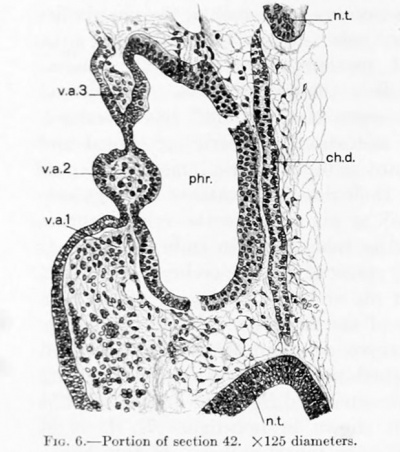
|
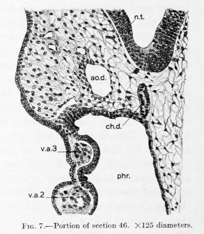
|
| Fig. 6. Portion of section 42. X125 diameters. | Fig. 7. Portion of section 46. X166 diameters. |
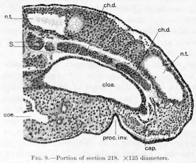
|

|
| Fig. 8. Portion of section 218. X125 diameters. | Fig. 9. Portion of section 222. X166 diameters. |
Watt describes what lie considers to be neurenteric canals in his twin embryos. They are situated ju.st dorsal to the cloaca and consist of a connecting rod of cells joining the chorda with the medullary tul)e dorsally and with the entoderm ventrally. This region in my specimen is cut through sagitlally, but as seen in text -figures K ;iii(l 0. no evidence of such a cord of cells is present.
The chorda dorsalis consists of polygonal and wedge-shaped cells, with large rounded nuclei and considerable granular cytoplasm. In places the cells are arranged radially about the center of the chord, while in other regions this arrangement is less distinct. ^Mitotic figures are numerous among the cells.
In the center of the chorda a fine lumen can be made out in certain sections (plate 4, fig. 3). This lumen is not continuous throughout, but is present in numerous places, each extending through but a few sections. In size it varies from 2 to 4 microns. That this discontinuous lumen of the chorda is normal in certain young stages of the human embryo seems to be well established, since it has been found by His'* in his embryo LI of 2.4 mm., by Eternod^ in three embryos of 1.3 mm., 2.11 mm., the third somewhat larger, and by Watt^* in twin embryos of 17-19 paired somites.
An ill-defined cuticular membrane surrounding the chorda dorsalis, such as has been described by Van den Broeck*^, is present, except where the chorda is fused to the entoderm of the pharynx.
Nephric System
In describing the Robert Meyer embryo of 23 somites (Thompson's embryo) Felix" states:
- "The pronephros is almost completely developed, so far at least as one may speak of its completion. It consists of a number of tubules and the primary excretory duct. There are in all seven tubules present, the most anterior of which is not united with the succeeding tubule .... The tubules 2-5 have fused so as to form a collecting duct. The tubules 5, G, and 7 are not yet united, but their union is imminent."
In my specimen I likewise find a .series of tubules representing the nephric
system. Although conditions on both sides of the embryo are not identical, for
the most part they are quite similar. The following description is based on their
arrangement of the left side and the terminology used is the same as employed
by Felix.
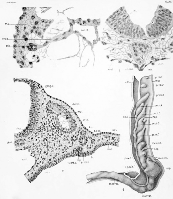
On the seven pronephric tubules which are present, the first two are rudimentary (plate 4, fig. 4). The first is represented by two or three small clusters
of cells at the level of the ninth body-segment. It is indistinct and indefinite and
its identity is determinable only by the position which the clusters of cells occupy
and by the fact that the cells are more or less isolated from the mesenchyma.
The second tubule is larger and more distinct. It is composed of a single cluster of cells which form a spherical mass. Its cells are isolated from the mesenchymal cells by a clear space. It also lies in the seventh body-segment. The remaining pronephric tubules are elongated gland-like tubes of epithelium, rather bulbous at their cephahc extremities and tapering caudally. Each in its course crosses the body-wall obliquely in a ventro-dorsal direction; thus in its upper part
each lies close to the mesodermal lining of the coelomic cavity, while below it comes in close relation with the ectoderm (plate 4, fig. 4). According to the description of Felix, each of these tubules represents two different portions of the pronephros. The bulbous portion is the inner pronephric chamber. It is connected to the mesothehum of the coelomic cavity by a slender strand of cells, the nephrostome, which joins it ventrally. The caudal portion of the tubule Felix terms the principal collecting tubule.
The third pronejihric tubule is the longest. It is rounded in cross-section and more regular in form than any of the succeeding tubules. The inner pronephric chamber is small cephalically, but is not connected to the mesodermal lining of the body-cavity by means of a nephrostome. A jnt on the mesodermal lining of the body-cavity opposite the tubule, however, probably represents a brokendown nephrostome. The principal collecting tubule extends caudally close to the ectoderm. It terminates in relation with the principal collecting tubule of the fourth pronephric tubule. Whether an actual fusion exists between the two collecting tulniles or whether they merely touch, I am unable to determine. The third pronephric tubule possesses a distinct lumen, which extends throughout most of its length. It lies in the tenth body segment, its collecting tubule extending into the eleventh segment.
The fourth tubule is almost the same shape and size as the third and is less
regular in form. Again a connecting nephrostome is lacking, although its position
is marked on the free surface of the mesothelium by a depression. The principal
collecting tubule ends in connection with that of the fifth tubule. The joined
distal ends of the collecting tubules give rise to the primary excretory duct. The
fourth tubule possesses a discontinuous lumen divided into two portions.
The fifth (plate 4, fig. 2), sixth, and seventh tubules differ from the third and fourth in possessing connecting nephrostomes. Each nephrostome begins as a
fiuinel-shaped opening on the coelomic wall. In no case, however, can a lumen
be traced into the tubule. Each nephrostome is joined to its inner pronephric
chamber by a strand of cells. Of these, that of the fifth tubule is extremely long
and band-like. The inner pronephric chambers are definite swellings situated
near the middle of the tubule. That of the seventh is the largest. The principal
collecting tubules are joined by the preceding ones as shown in plate 4, figure 4.
Lumens are present only in the inner pronephric chambers. The fifth and sixth
pronephric tubules are situated in the twelfth body-segment, while the seventh
is found in the thirteenth.
Histologically the tubules in all parts arc made up of polygonal or columnar cells, with rounded or elongated nuclei. Where a lumen is present the cells are arranged radially about it, but in other places there seems to be no definite arrangement.
Beyond the seventh pronephric tubule and apparently continuous with it is a large rounded cord of cells which extentls through the remainder of the embryo. This cord possesses a number of irregular swellings, the mesonephric vesicles. These vary in size and shape and arc not so definitely marked off from one another as shown by Felix" and Watt"; 16 to 18 may be counted (plate 4, fig. 4); they possess nephrostomes connecting them with the coelomic mesothelium similar to those of the pronephric tubules.
The primary excretory duct occupies a position just dorsal to the nephrogenic cord. In its course caudally it lies close to the ectoderm. Felix was unable to determine whether it develojied from the ectoderm or whether it arose independently in the mesoderm, but he doubts that the ectoderm has any participation in
its development. Watt^* also was unable to determine definitely its mode of formation. The caudal end of the primary excretory duct, the point at which its formation is supposed to be taking place, is in my embryo cut sagittally. It is, however, indefinite. Just before its termination the duct is seen lying close to the ectoderm in the mesenchyma. The mesenchyma possesses several mitotic figures in the region in which it terminates. While I am incUned to favor P'elix's view of a mesenchymal origin, the evidence found is not convincing.
Vascular System
Heart
The heart lies in that portion of the body-cavity which is bounded by the pharynx above, the fore-gut behind, the anterior body-wall in front, and the transverse septum below. It is still a simple tube and viewed from in front is roughly the shape of the letter U. It is placed so that the loop of the U is directed toward and lies in the right side of the pericardial cavity. The limbs of the U are turned to the left, but upon reaching the body-wall of the left side turn dorsalward at an angle of almost 90 degrees. The upper limb then bends sharply cephalad, joins immediately the pericardial wall, and passes into the ventral aorta. The lower limb bends medially and joins the pericardial wall as it passes into the sinus venosus. The ends of the heart-tube are therefore fixed to the bodj^-wall, but the remainder of the heart lies free within the cavity.
An examination of the heart-tube shows that it is not everywhere of the same caliber, but presents certain exjianded portions separated by more or less definite constrictions. Beginning with the venous end of the heart, the sums venosus passes into the atrium with but a sUght constriction. Figure 2, plate 1, shows that portion of the heart which I regard as the atrium. As seen from behind (plate 3, fig. 1), it presents a V-shaped bend, the apex of which is pointed toward the left, the limbs lying in a horizontal plane. On its upper surface is a distinct irregular projection, as to the significance of which I am in doubt.
The atrial portion of the heart passes into the ventricular i)ortion with only a sUght constriction, the atrio-ventricular canal. The ventricle is much enlarged, having a transverse diameter which is greater than that of any other portion of the heart. It fills up the entire lower right-hand portion of the pericardial cavity. Its cephahc end is bounded by a shallow constriction, in front of which is the bulbus cordis.
The bulbus cordis is large where it is attached to the ventricle, but gradually becomes narrowed towards its cephalic end. It is directed downwards and towards the left. As described by Watt and others, it reaches farther cephalad than any other portion of the heart. It becomes continuous with the truncus arteriosus, a short, narrow portion of the heart-tube which is directed dorsally and towards the left. The truncus arteriosus bends sharply upward and joins the ventral aorta.
The cavity of the heart approximately follows the center of the heart-tube. Like the outside of the heart, it also is not of uniform diameter, but presents swelUngs and constrictions, as shown in plate 3, figure 2. The sinus venosus passes to the left as an irregular tube. It is marked off from the atrium by a sUght constriction. At the apex of the atrial bend the cavity enlarges considerably. On the superior surface of this enlargement is a crescentic projection, the concavity of which is directed medially. This extends into the above-described j^rojection of the atrium as seen from the surface.
Between the atrium and ventricle is a long, narrow portion of heart-cavity, the atrio-ventricular canal. The difference in size between this narrow portion and that of the adjacent swellings is greater than that between the same portions of the heart as seen from its surface. The cavities of the ventricle and bulbus cordis are again enlarged and separated from one another by a rather long constriction portion. This is again much narrower proportionally than the same constriction seen from the surface of the heart. In cross-section the cavities of the ventricle and bulb, particularly the former, are triangular. In the truncus arteriosus the cavity is again small and irregular. Cephalad it gradually widens out as it becomes the ventral aorta.
The endothehum of the heart is everywhere a syncytium of one layer of cells. Its nuclei are oval in shape and coarseh' granular, while the cytoplasm stands out sharply in contrast to the coagulum without. In the bulb and ventricle, at the angles of the cavity as seen in cross-section, the endothelium sends out platelike processes of cells. These give to the various portions of the endothelial heart a stellate appearance. In some instances these processes extend to the myocardial layer and apparently fuse with it. One such process is shown in plate 4, figure 1. It i)asses off from the endothelial tube of the heart as a solid cord of cells. It extends entirely across the space between the endothelial and myocardial layers and terminates in a bulbous expansion in contact with the myocardium. In the expanded end is seen a distinct mitotic figure. The significance of processes of this kind can not be doubted. They represent the earliest lieginnings of the so-called "heart sinusoids," which in embryos a little older are numerous. By their growth and anastomoses they give rise to the trabecuke of the heart.
In the atrium, with the exception of the above-described atrial projection, the endothelium lies in contact with the myocardium, there being no intervening space such as is found in the remainder of the heart-tube. This relation of the endothelium to the myocardium in the atrium has been noted by His'*. The same condition has lieen described by Mall", who states:
"This Jirrangement is so pronounced in the early heart that it affords a way by which we may detomiine with precision the exact portion of the heart tube from which the atrium arises."
No mention, however, is made by Mall, or by \N'att", who describes a similar
arrangement, of that portion of the atrium which projects upward and in which
the endothelium is not closely applied to the myocardium.
The outer layer of the heart-tube, the so-called myo-epicardial layer, is composed of several layers of cells, the thickness varying in different portions of the heart. As Tandler" has shown in an embryo of 3.5 mm. (embryo Hal, Institute of Anatomy, Vienna), these cells form a distinct syncytium. The myo-epicardium is directly continuous at the venous and aortic ends of the heart with the transverse septum and Uning of the body-cavity, respectively, and it is by means of these attachments that the heart is held in place.
In that portion of the atrium where the myo-epicardium lies in contact with the endothehum, its cells are closely appHed to one another. Nuclei are crowded and intercellular spaces are small. The myo-epicardium of the atrio-ventricular canal is somewhat thicker than that of the atrium, and its cells are more widely separated. Numerous small protoplasmic processes of the cells form a network, the meshes of which are fiUed with delicate fibrils. In the ventricle and bulbus cordis the myo-epicardium is still thicker, but in the truncus arteriosus it again becomes thin.
In describing the 3.5 mm. embryo, Tandler" says:
"The myo-epicardial mantle differentiates to the extent that in the region of the ventricular loop and in the bulbus its superficial layer is formed by a continuous row of cells, the epicardium, while on the atrium and sinus, so far as the latter has a free surface, no such dififerentiation can be said to exist."
In my embryo I find that the outside layer of cells of the myo-epicardium have not yet become flattened and detached from the underlying cells, as shown by Tandler in his figure 377, but over the entire surface of the heart the}' are arranged in a distinct laj'er (plate 4, fig. 1). As seen in cross-section, they are in places cubical and closelj' packed, while in other places they are more rounded and farther spread apart. I am also unable to find any differentiation of the myocardium proper into an inner spongj' portion and an outer cortical portion, as Tandler describes for the 3.5 mm. specimen.
The broad space which exists between the myo-epicardial and endotheUal layers, according to Tandler, is filled during life with serous fluid, since it is occupied in section by a clot-Uke fibrous mass which is entirely destitute of cells and stains feebly with the hemotoxj'hn. In my specimen I find a similar clot-like mass. The small, delicate fibrils form an anastomosing plexus, the meshes of which are empty. For the most part these fibrils extend radially from the endothelium to the myocardium, to both of which thej' gain attachment. They radiate out particularly from the plate-like processes of the endothehum, making it impossible to determine in every case where the endothelium leaves off and where the fibrin begins. It seems probable that the deUcate fibrils found in the myocardium are Oue to a similar coagulation of serous fluid.
Mitoses within the tissue of the heart are few. Occasionally one may be observed in the endothelium, particularly in the cells of its plate-like processes.
In tho myoepicardium, witli the exception of that of the sinus venosus, they are extremely rare. In the sinus venosus, and particularly in those portions of the mesot helium of the body-cavity and transverse septum which are directly continuous with the myo-epioardium, mitotic figures arc comparatively numerous. Tliis fintUng would seem to indicate that the multiphcation of cells of the myoepicardium of the heart takes place at this stage principally in the region of tlie sinus venosus.
Veins
Vena Cardinalis Anterior
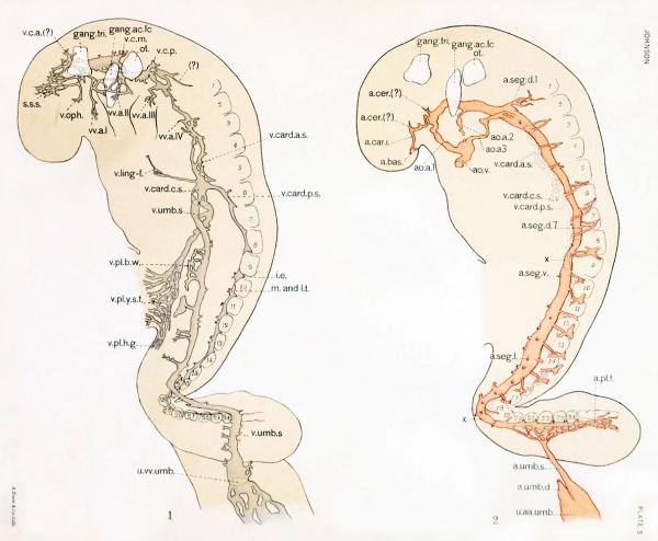
The vena cardinalis anterior (plate o, fig. 1) draws its blood principally from the region of the brain. In Ingalls's embryo this vein begins at the junction of two veins which course caudally from the region of the prosencephalon. Ingalls, basing his interpretations upon the work of Mall", regards the dorsal of these as the source of the future sinus sagittalis sujierior; the ventral one, the vena ojihthalmicus. In my specimen the region drained by the two above-mentioned veins is occupied by a venous plexus. One tributary, which extends upward from the region of the optic vesicle, may already be identified with reasonable certainty as the ophthalmic vein. The much more extensive plexus above probably gives rise to the embryonic superior sagittal sinus. The tributaries of the abovedescribed plexus come together medial to and behind the trigeminal ganglion, where they form an enlarged venous sinus, between the trigeminal ganglion in front and the acustico-facial ganglion behind. From this the anterior cardinal vein passes caudally by two main channels, the dorsal of which is situated above the origin of the acustico-facial ganglion, while the ventral one is placed medial to it. Passing the interspace between the acustico-facial ganglion and otocyst, these branches unite again medially to the otocyst, forming another enlarged portion of the vein. Caudal to the otocyst it divides into three smaller veins, which soon come together again. Opposite the first somite the anterior cardinal vein becomes very .small in diameter, but gradually becomes larger when traced still farther caudally. From the second segment to its termination, the vena cardinalis anterior is represented by two small veins which are closely related to one another. In several places, however, they unite to form a single vessel. At the level of the fifth body-segment the vena cardinalis anterior enters the vena cardinalis communis (duct of Cuvier).
In its course the vena cardinalis anterior receives tributaries on both its dorsal and ventral walls. On the dorsal, the first is found in the region of the trigeminal ganghon and proceeds from the direction of the mesencephalon to reach the anterior cardinal vein at the posterior border of that ganglion. The second lies just in front of the otocyst close to the rhombencephalon. The position which it occupies (just in front of the otocyst) indicates that it is the same vein which Mall" describes lus the vena cerebri media. Behind the otocyst is a small stump of a vessel which could not be traced far dorsally. Owing to its position. just behind the otocyst it becomes evident that this vein must be identical with the one occupying a similar position in Ingiills's embryo mih! which i'Aan.s" has interpreted to be the vena cerebri posterior. Just caudal to this vein is a longer vein, which, arising in the region of the first segment and extending anteriorly and ventrally, joins the anterior cardinal vein at the point where the latter is broken up into two portions. I am in doubt concerning its identity. Several small dorsal tributaries of varying size are received throughout the remainder of the vena cardinalis anterior. They include the lateral loops of the second, third, and fourth dorsal segmental arteries, which are described below.
The ventral tributaries are more numerous than the dorsal. They may be
described as belonging to the different visceral arches. One arises in the mandibular arch close to the mouth, passes dorsalwards, and unites with a network
of small veins. The blood from this tributary may reach the anterior cardinal
vein, either in the region of the trigeminal or of the acustico-facial ganglion.
The venous tributaries of the second arch do not arise as far down as those
of the mandibular arch. They form a plexus which lies in close relation to the
facial nerve. One tributary passes medially to the ganglion, while the others are
laterally situated. There is thus established about the acustico-facial gangUon a
venous ring. The plexus anastomoses with that of the first arch.
In the upper part of the third arch are found three small tributaries, which
unite and reach the anterior cardinal vein as a single vessel. No others are found
in this visceral arch. Farther caudally, opposite the second and third somites,
several small veins unite with the anterior cardinal. They probably represent
similar tributaries from the fourth arch.
At the point at which the anterior cardinal veins empty into the common cardinal there is received, on the ventral side, a long, slender vein (vena linguofacialis). The smallest tributaries of this vein may be traced as far as the third visceral arch. Uniting, these tributaries form the vein which proceeds caudally and dorsally. In its course it passes in the antero-lateral body-wall over the heart. A similar vein has been described by Ingalls'*, as follows:
- "Am .\nfang des Ductus Cuvieri miinden in ihn auf jeder Seite je ein vou der vorderen Bauchwand kommendes Gefass. Auf der rechten Seite ist dies besonders gro.ss,
es lasst sich ventralwarts bis in die Nahe des Ursprungs der ersten Aortenbogen verfolgen and weiter Kaudahvarts bis daliin, wo die ersten Kiemenbogen mit der vorderen Korperwand verschuiolzen sind, um sich schlies'^lich in dem ersten Bogen zu verlieren."
The same vein had been found by Salzer^" in embryos of the guinea-pig and later by Grosser'* in bat embryos. Lewis^* describes it in the pig embryo under
the term "transverse vein," and later^^ discussed its origin and fate in rabbit and human embryos. Apparently the first reference to this vein, which is now
recognized as of constant occurrence and fundamental morphological importance, was made by Phisalix, as Dr. Lewis has pointed out to me. Phisalix", in 1888, showed it clearly in a figure of a 10 mm. human embryo and described it as follows:
- "Entre la veine jugulaire et la veine cardinale se trouve une vaste poche dont le sang s'ecoule par les canaiix de C'uvier .... Kn avant et au-dessus, chacune de ces poches regoit des veinules qui acconipagnent le nerf hypoglosse et qui ^^ennent de la base des arcs branehiaux."
In injections of the veins of pig embryos, Smith^' shows definite anastomoses between the Hnfjuo-facial vein and the venous plexuses of the visceral arches. The anastomosis between this vein and the plexus of the first visceral arch is apparently already formed in Ingalls's 4.9 mm. human embryo. In my embryo I have been unable to find an anastomosis, but the direction which the vein takes (plate 5, fig. 1) seems to indicate the possibility that these two sets of tributaries might soon unite.
Vena Cardinalis Posterior
The blood from the tail and lower half of the embryo is carried to the vena cardinalis communis by means of the vena cardinalis posterior. This arises in the tail of the emliryo in the region of the unsegmented mesodermal plates as a slender irregular vessel. In its course to the bend in the back it passes along the ventrolateral border of the somites, with which it lies in contact. In this part of its course, owing to the plane of sectioning, it is difficult to trace its capillary connections, but in places there is evidence that connections similar to those above the nineteenth somite exist below it.
For the most part that portion of the i)osterior cardinal vein from the bend in the back to the conmion cardinal stands out quite sharply. In general it courses along the ventro-lateral border of the somites, lying between them and
the pronephros or mesonephros. Opposite the somites of the seventh to eleventh
segments the posterior cardinal vein becomes very difficult to follow. It is represented by a slender and apparently solid cord of endothelial cells. In the
intersegmental spaces the vessel becomes larger and usually contains a number
of blood-cells (plate 5, fig. 1). Opposite the ninth somite (and again the tenth)
the existence of the vessel becomes doubtful, owing to the similarity of endothelial
to mesenchymal cells. In the corresponding region on the right the continuity
of the posterior cardinal vein is even more doubtful. Whether this is due to a
closing-uj) of a once continuous posterior cardinal vein or whether it represents
an incompletely developed vein is impossible to determine from the sections.
According to Evans*, the posterior cardinal veins first apjiear in human embryos
possessing from 15 to 23 somites. He states:
- "It is possible that lateral loops of the dorsal segmental arteries are instrumental in the formation of these veins, as in the case of the anterior cardinals. This nietliotl of
formation of the posterior cardinal veins appears fimdamental. RafTaele (1892) and Hoffman (1S93) describe it for selacliian embryos and (irafe (1905) and the writer have indicated it in the case of the chick."
It is probable, therefore, that in my embryo the above-mentioned portions
of the posterior cardinal veins are still in the formative stage, particularly since
the intersegmental portions of the vein, to which are joined the lateral loops of
the segmental arteries, are more (h'linitely marked than the segmental portions.
From the interspace between the seventeenth and eighteenth segments to its termination, the posterior cardinal vein receives its blood principally from the small lateral loops of the dorsal segmental arteries, as has been described by Evans*.
In addition to these tributaries, connections can be made out in several places between the posterior cardinal vein and the lateral branches of the aorta, but these connections are not so distinct as the branches from the dorsal segmental arteries. In the regions of the pronephros and mesonephric vesicles small tributaries are found arranged in pairs, one lying on either side of these organs; both pass dorsally to join the posterior cardinal vein. The lateral tributaries pass, in the region of the pronephros, between the inner pronephric chambers of one pronephric tubule and the principal collecting tubule of the preceding one. Farther down, in the region of the mesonephros, they pass between the mesonephric vesicles and the primary excretory duct.
Evans* shows similar vessels in a reconstruction of the 23-somite embryo of Robert Meyer. He also indicates a longitudinal vein which connects the peripheral ends of the medial tributaries and another similarly connecting the lateral tributaries; these he terms the medial and lateral subcardinal veins, respectively. I have been unable to make out continuous longitudinal connections in my specimen, but indications of them are apparent on a few of the medial vessels. I find, however, connections between the medial tributary and the lateral segmental arteries, such as Grafe'^ has shown in the chick and Evan-s* notes in the Robert Meyer specimen. It is interesting to note that the two above-described tributaries of the posterior cardinal vein are not located with reference to the segments, but (as Evans and others have described for the lateral segmental arteries) they correspond quite closely in number and position with the pronephric tubules, where the latter are present. Below the pronephric tubules they are arranged with reference to the mesonephric vesicles.
Vena Cardinalis Communis
The common cardinal vein (duct of Cuvier) is a short, flattened vessel which lies within the sixth body segment. It receives both the anterior and posterior cardinal veins. It is directed caudaUj^ in the lateral body-wall and, breaking up into three portions, joins the vena umbiliealis at the point where this vem enters the transverse septum (plate 5, fig. 1, and plate 6, fig. 1).
Venae Umbilicales
The umbilical veins begin at the distal end of the body-stalk by the union of several large veins which drain the chorion and its villi. At first they form a single vessel, which soon, however, breaks up into a plexiform arrangement of large veins (plate 5, fig. 1). These reunite to form a single large vein, which again divides to form two smaller vessels, the right and left umbilical veins. Immediately upon separating they pass to the outer border of the umbilical stalk, one on either side, and enter the body-wall.
In the beginning of its course the right vein is very small. Soon, however, it increases in size and in the remainder of its course it is similar to and about as large as the left umbilical vein, which is quite uniform throughout. Each umbilical vein, throughout its entire course from the body-stalk to the septum transversum, lies within the body-wall, situated in this at about the junction of its middle and distal thirds. Each receives from tlio Viodj--wall tributaries coiniiif? from both dorsal and ventral directions. The tributaries which reach the umbilical vein on its ventral 'wall arise for the most part within villus-like processes of the body-wall. On the left side, one of these (found at the level of the tenth and eleventh body-segments) is even larger in cross-section than the main stem of the vein itself and forms a venous sinus in the villus (plate 5, fig. 1.) It is drained by a relatively small vessel. Above this are a number of tributaries which drain a longitudinal vessel situated in line with the venous sinus below. Concerning the significance of these vessels I am in doubt, but beheve them to be either the remnants of the plexus from which the umbilical vein has developed (Evans*) or the beginning of the anterior body-wall plexus (Smith^').
Opposite the seventh somite the left umbilical vein receives a branch from the vitelUne vein, which lies ventral and caudal to the main junction of these vessels. Approximately at the level of the interspace between the sixth and seventh body-segments, the left umbilical vein receives from above the vena cardinalis communis, entering by three distinct tributaries (plate 5, fig. 1), as described above. Turning sharply medially into the septum transversum, it unites with the vitelline vein (plate 6, fig. 1), and with it forms the left vitello-umbilical trunk. This trunk, which is represented by one main vessel and two smaller ones, passes medial to join the sinus venosus.
The right umbilical vein likewise receives tributaries from the body-wall all along its course and at its most cephalic point receives the right common cardinal vein. It is united to only a portion of the vitelline vein and at but one point. The vitello-umbilical trunk is represented on this side by a network of smaller veins which connect it w'ith the sinus venosus.
Venae Vitellinae
The vitelline veins arise on the surface of the yolk-sac from the yolk-sac plexus. Two principal veins, the right and left, are formed on the yolk-stalk by the convergence of numerous tributaries. These course cephalad, one on either side, and enter the septum transversum (plate 6, fig. 1). They pass dorsally along the sides of the hepatic diverticulum, lying quite close to it and the wall of the fore-gut. Several small tributaries proceeding from the mesenchyma surrounding the fore-gut are received by them from above. Each breaks up into several branches, which form a plexus within the transverse septum. A minute commissural branch connecting the veins of both sides is found in the notch between the hepatic diverticulum and the fore-gut wall. The two coiniections of the left vitelline vein with the left umbilical vein have already been described. All the blood carried by the left vein, except that which may cross over to the opposite side in the small comniissural branch, joins (hat of the umbilical vein before reaching the sinus venosus. Near its termination the right vitelline vein breaks up to form a plexus, the branches of which diverge. Some of these pass directly into the sinus venosus, while others join the umbilical vein to form a plexus representing the right vitello-umbilical trunk. Only a part of the blood which is carried by the right vitelline vein, therefore, pjusses through the right vitello-umbilical trunk.
Over the yolk-stalk the tributaries of the vitelline veins form a network of small vessels. These reach dorsally as far as the middle of what might be outlined as the gut. Extending along the gut-wall, therefore, are a number of small longitudinal coursing veins (plate o, fig. 1). They can be traced caudally along
the hind-gut for some little distance below the hind-gut portal. The significance
of this plexus I have not been able to determine definitely, but I judge that it will
ultimately give rise to the inferior mesenteric vein.
Sinous Venosus
The sinus venosus is situated within the substance of the septum transversum, ventral to the gastric region of the fore-gut and cephalad to the tip of the hepatic diverticulum. It is a broad, irregular vessel (text-fig. 4 and plate 5, fig. 1), much flattened dorso-ventrally. On its left border it receives three vessels which repre.sent the left vitello-umbilical trunk. On its right it receives three small branches from the right vitello-umbilical trunk and as many more directly from the right vitelline vein. The sinus venosus curves ventrally and to the left and, becoming a more rounded vessel, passes out of the transverse septum into the atrium of the heart.
Arteries
Aorta Ventralis
The ventral aorta (plate 2, fig. 3, and jjlate 3. figs. 3 and 4), the direct continuation of the truncus arteriosus, is an unpaired median vessel. It is situated just ventral to the thyroid diverticuhun. with which it lies in contact. It at once breaks up into the aortic arches.
Aortic Arches
Three pairs of aortic arches are present (plate 5, fig. 2). Of these the first is by far the largest. Each vessel begins at the anterior extremity of the ventral aorta and extends anteriorly and dorsalward in the first visceral arch, just cephalad to the first pharyngeal pouch. Reaching the upper extremity of the arch, it joins with the dorsal aorta. Both first arches are distinctly patent throughout.
The second and third aortic arches are smaller and less distinct vessels, lying within the second and third arches respectively. The vessels of these arches vary in size and distinctness in different regions. In certain places they become so small that a lumen is no longer discernible, and it becomes impossible to determine whether the vessels are continuous or not. It is very probable, however, that connections do exist at these places, but, owing to the great similarity between the endothelial cells and those of the surrounding mesenchyma. the former can not be traced through with any degree of certainty. On the left side the second arch
shows two such doubtful interruptions, one at the jwint where it leaves the ventral aorta, the other where it joins the dorsal aorta. Between the two breaks a distinct vessel is present. The third arch on the left side arises from the dorsal aorta as a small plexus and extends as a distinct vessel halfway down to the ventral aorta. Here it disappears for several sections, but soon reappears as an apparently solid string of endothelial cells. This cord when traced downward is found to connect with the ventral aorta. On the right .side the second and third aortic arches likewise can not be traced from dorsal to ventral aorta. The apparently absent
portions of both of these arclies are near the ventral aorta.
As shown in plate 5, figure 2, there is on the left side a small arterial twig which branches off from the dorsal aorta just behind the third arch. The significance of this is uncertain, but from its position it seems (juite probable that it may be the beginning of a fourth aortic arch.
Whether the second and third arches are not yet comi)letely developed or whether their lumens have become secondarily occluded I am unable to determine. It would seem, however, that the former is the more probable, even though in the somewhat younger embryos of Watt and Van den Broeck,^ and in Thompson's embryo as described by Felix, the second arch is complete. The beginning third arch (and the probable beginning fourth) indicates that in this respect my specimen is older than any of the above-mentioned embryos. The incomplete second arch may be regarded as having been slightly retarded in its development.
Aortae Dorsales
The dorsal aortae are two large vessels which extend from about the level of the anterior end of the chorda dorsalis to within a very short distance from the tip of the tail. Between the eighth and nineteenth segments the dorsal aortse are fused together in the mid-line, forming a single median vessel; elsewhere two distinct vessels are apparent. Where two vessels are present they lie one on either side of and slightly ventral to the notochord; where single it lies directly ventral to the notochord. Throughout their entire courses, whether paired or single, the dorsal aortae lie just dorsal to the digestive tube.
The dorsal aortae, when traced from their anterior to their posterior extremities, continually change in shape and size. The median dorsal aorta is smallest in cross-sectional area at about the level of the eleventh segment and largest at the level of the fourteenth. The vascular bed (cross-sectional area) at this level is even larger than that of the paired dorsal aortas combined. The endothelium of the dorsal aortae is distinct throughout. In the region of the fourteenth segment it forms an incomplete septum, undoubtedly the remains of the originally fused medial walls of the ])aired vessels, which have not as yet disappeared.
Branches of the Dorsal Aortae
Anterior Branches. -At the i)oint where the dorsal aorta and the first aortic arch join, two small arteries (plate 5, fig. 2) are given off from the dorsal wall of the dorsal aorta. The one situated more caudally is the smaller of the two. It extends dorsally and medially and terminates in the region of the posterior end of the prosencephalon, a short distance behind the anterior end of the notochord. I am unable from the specimen or from other descriptions to identify this vessel with any degree of certainty, but presume that it gives rise to one of the cerebral arteries. The anterior branch is larger. It extends medially and anteriorly and comes to lie close to the side-wall of the prosencephalon. Here it divides into two branches, one of which extends forward on the ventral wall of the prosencephalon, while the other passes medially along its ventral wall. The latter branch terminates near the mid-line of the embryo not far from its fellow of the opposite side. In addition, the anterior branch gives off along its course two or three small arterial twigs. These pass dorsally along the wall of the brain, and probably represent the beginning of the cerebral arteries.
Dorsal Segmental Arteries. - The dorsal segmental arteries are represented by
24 paired vessels. Although for the most part they are similar in position and
distribution, they vary greatly in size and in the distinctness with which they can
be traced. The following description refers only to those of the left side.
In his account of these vessels of the Robert Meyer embryo Xo. 300, Evans states* :
- "At this stage the dorsal segmental vessels form in the tissue of the intersomitic clefts large, well-marked vascular arches or loops, one limb of which is against the neural tube, while the other joins the cardinal vein."
Evans had previously shown similar loops in chick embryos.
In my embryo I find that loops are present or indicated in case of all of the upper 18 dorsal segmental arteries except the first. Two such typical vessels are shown in plate 6, figure 2. Each dorsal segiaental artery extends dorsally and laterally from the aorta. When it reaches a point about halfway up the medullary
tube it branches, sending a short branch mediallly and a longer one laterally. The
lateral branch extends through the intersomitic cleft and reaches the posterior
cardinal vein as described by Evans. The medial one, I find, soon divides into two
distinct smaller branches, a dorsal and a ventral. These extend along the wall of
the medullary tube and tend, with their fellows of the opposite side, to encircle it.
The first dorsal segmental artery has a relatively extensive origin from the
dorsal aorta. It extends dorsalward between the first and second somites, where
it breaks up into several branches. One of these extends for a short distance
anteriorly, while others extend caudally toward the second dorsal segmental.
A connection between the two, however, is apparently not yet formed. I am
also unable to trace a connection between this artery and the anterior cardinal vein. In Ingalls's specimen of 4.9 mm. the first dorsal segmental artery (known
also as the hypoglossus artery) likewise sends branches both anteriorly and
posteriorly. The posterior branch, however, has joined the second dorsal segmental artery, while the anterior is much longer and is easily recognized as the
arteria vertebralis. In my specimen, therefore, one sees the very beginning of the formation of the vertebral artery.
The second and third dorsal segmental arteries show only lateral and dorsal
branches, while for the fourth only pieces of a typical dorsal segmental artery
could be identified with certainty in the sections. It seems improbable that this
and other similar vessels are actually incomplete, more probable that they are
complete and that in places are so small and indistinctly differentiated from the
mesenchyma th^^: the connecting portions have been overlooked. The lateral
branches of the second, third, and fourth dorsal segmental arteries unite with the
anterior cardinal vein; those of the remaining dorsal segmental arteries join the posterior cardinall vein. The Hfth and sixth dorsal segmental arteries are shown
in white (i, figure 2. The seventh and eigeth are similar to the fifth and sixth; in addition the eighth presents a bulbous swelling which lies against the lateral
wall of the medullary tube. The ninth is almost typical, but its ventral branch is either lacking or indistinguishable. The tenth to seventeenth show bulbous swellings (similar to that of the eighth) of various size extending longitudinally along the neural tube. At some jilaces these swellings extend toward one another and probably form the longitudinal anastomoses along the neural tube, which Felix'"
has indicated are present in the Robert Meyer embryo Xo. 300. Evans" has shown
that such anastomoses exist in the form of a distinct plexus in the chick embryos.
In my specimen anastomoses very probably are present, for what appear to be networks of endothelial cells connecting adjacent segmental arteries are distinguishable
in many places.
The eighteenth to twenty-fourth dorsal segmental arteries seem to be less well developed. They can only be followed with difficulty, owing to their smallness and to the plane in which the sections are cut. Their origins from the aorta. however, are very apparent.
Ventral Segmental Arteries. - The ventral segmental arteries are paired vessels, but they are found only in the lower segments of the body and their segmental arrangement is not so definite as that of the dorsal branches. According to Evans* there is originally a ventral artery for each segment, but those of the upper body-segments degenerate very early. The upi^er ones together constitute a row of vitelline arteries, which later, by fusion of the individuals of certain pairs, give rise to the unpaired median vessels of the adult.
In my specimen there are 19 to 20 pairs of ventral segmental arteries in all, including those which go into the formation of the umbilical arteries and which
should undoubtedly be classified as ventral segmentals. They extend from the
seventh segment to the tail. The largest ones are placed opposite the seventeenth
and eighteenth somites. All of the ventral segmental arteries pass ventrally along
the wall of the digestive tract. Anastomoses, such as Felix has shown, can be
made out in certain places. Most of the branches above the nineteenth segment
can be traced to the yolk stalk and sac, where they become larger and enter into
an extensive plexus. Those below the nineteenth segment go into the formation
of the umbilical arteries, as described below.
Lateral Segmental Arteries. - Lateral .segmental arteries are found opposite the twelfth to nineteenth segments. They are small vessels, directed laterally at right angles to the longitudinal exis of the aorta. They can be traced as far out as the nephric system. In several jilaces connections can be observed between them and the termination of the medial tributary of the posterior cardinal vein (the beginning median subcardinal veins as described by Evans). I have been unable to determine whether or not lateral segmental vessels exist below the nineteenth .segment, owing to the plane of section and to their indefiniteness.
Terminal Branches. - The dorsal aortae terminate in the tail of the embryo by breaking up into distinct networks of small vessels. These plexuses probably represent the caudal arteries in their earliest stage, but as yet they can not be said to exist as definite arteries. I have referred to one of them in plate 5, figure 2, as the "arterial plexus of the tail."
Arteriae Umbilicales. - The lunbilical arteries are formed by the union of the lower ventral segmental arteries, including all those caudal to the twentieth segment. Apparently one or two small capillary twigs from the tail plexus also enters into its formation. Anastomoses between the individual roots of the artery are apparent, thus giving rise to a network. In plate 5, figure 2, the network is
represented diagrammatically, since I found it unprofitable to attempt to plot them with any degree of accuracy. In the first part of their course, where they lie on either side of the allantoic duct, the umbilical arteries are small and indistinct. Soon, however, they rapidly enlarge and fuse together to form a large trunk within the body-stalk. Reaching the chorion, the single umbilical artery breaks up into a number of branches, which, after repeated division, terminate as capillaries in the substance of the villi. The relation of the allantoic duct to the fork formed by the fusing umbilical arteries has been described above.
Coelom
The coelom is represented by a continuous elongated cavity which is in wide communication with the extra-embrj-onic coelom. Already it can be divided into two distinct parts, the pericardial and the pleuro-peritoneal cavities. The first of these surrounds the heart, except where the heart is attached by means of its sinus venosus behind and by its truncus arteriosus above.
Pericardial Cavity
The pericardial cavity reaches its highest point in the region of the bulbus cordis. Its form is shown in plate 6, figures 3 and 4. It is bounded dorsally by the pharynx and septum transversiun, ventrally and laterally by the thin bodywall. Its floor is formed by the septum transversum. The floor is deficient on either side dorsally where the transverse septum is not yet complete, and in the space between it and the posterior body-wall the pericardial cavity establishes its commimication on either side with the pleuro-peritoneal cavity which lies below.
Pleuro Peritoneal Cavity
The pleuro-peritoneal cavity is divisible into two portions, an upper and a lower. The upper is formed by two narrow limbs which unite below the yolkstalk to form the single lower ])ortion. The limbs of the upper portion join the pericardial cavity high up on its dorsal surface. At first they are directed dorsally and caudalward, but very soon bend directly caudalward. As viewed from behind, their median borders are not straight, but each presents two curves, the concavities of which are directed medially. Fitting into the spaces formed by these curves, as shown in plate 6, figure 4, is the lung diverticulum above and the hepatic diverticulum below. That portion of the digestive tube which I have considered as the gastric region lies opposite the constricted area between the two enlarged spaces.
Caudally the two limbs terminate by uniting below the yolk-stalk. Slightly above this level the pleuro-peritoneal cavity joins the cxoca?lomic cavity on either side and in front where the anterior body-wall is deficient; that is, following the line of reflection of the amnion (plate 6, fig. 3). That portion of the exocoelomic cavity which lies in front bridges across the space between the two above-described limbs. The bridge Hes just above the yolk-stalk, while lying between it and the pericardial cavity are the amnion, the amnionic cavity, and the anterior pericardial wall.
The lower portion of the pleuro-peritoneal cavity lies in relation to the liiudgut dorsally and the posterior surface of the yolk-sac ventrally. On either side it is in wide communication with the exocoelomic cavity. As seen in plate 6, figure 3. its anterior surface is irregularly ])itted, the cast of the irregular surface of the yolk-sac. Viewed from behind (])late (i. fig. 4), is a deep groove which marks the position of the hind-gut and dorsal mesentery. At the dorsal bend in the back of the embryo the ccclomic cavity continues caudally by means of two prolongations (plate 1, fig. 2. and plate 6, fig. 4). These lie laterally to the hind-gut and cloaca. They extend about as far caudally as the cloacal membrane. It is to be noted that these are not united together ventrally; consequently there exists in this portion of the embryo a ventral mesentery as well as a dorsal one.
Mesothelium
The body-cavity is everywhere lined with a mcsothelium which varies in character and thickness in different regions. As a rule, how^ever, it may be said that the visceral layer is thicker than the parietal. This is not entirely true regarding that portion of the ])arictal jiericardium which lies next to the fore-gut, for this is thicker, being composed of two to three layers of cubical cells. Since, however, this layer lies next to the fore-gut, it is in reality visceral, so that its being thick is not actually at variance with the general rule. The parietal pericardium elsewhere, i. e., lining the body-wall and the pericardial surface of the septum transversimi, is thin, being composed of a single layer of cubical cells. The visceral pericardium, the previously described peicardiuni. is formed by a single layer of cubical or rounded cells.
The parietal peritoneum, with the exception of that covering the peritoneal surface of the septum transversum, is thin and formed by a single layer of flattened cells. Where this becomes continuous with the visceral peritoneum (that is, on the dorsal mesenterjO it gradually becomes thicker. The inferior surface of the ."septum transversum po.ssesses a comjiaratively thick e])ithelium. being composed of three to four layers of cells. The visceral peritoneum is thick over the fore-gut and yolk-stalk, somewhat thinner over the hind-gut. and Ihiimest over the yolksac, where in most places it becomes lost as a distinct layer.
The lining of the extra-embryonic cavity, i. e., the inner lining of the chorionic vesicle, is not in the form of a distinct cell layer, but apjiears to be made up of uncovered mesenchyma. Mitotic figures are fovuid throughout all parts of the visceral Mud parietal epitheliuin, excejil in tlu> epicardium.
Septum Transversum
The septum transversum is a thick plate of mesenchyma which is Uned on either side with the mesotheHum of the ccelomic cavity. It divides off the pericardial from the pleuro-peritoneal cavity, except posteriorly and laterally, where it is incomplete. It is placed across the body at the level of the venous end of the heart and liver diverticulum and is directed obUquely from in front dorsallj' and cephalad. As near as can be determined, it is placed o]>posite the fourth and fifth body-somites. With reference to Mall's" schema (Mall's fig. 400), it may be said that in position the transverse septum of my embryo lies dorsally at a corresponding position which he has shown for embryos of 2 and 4 mm., and is directed at an angle which would pass some place between those of the 2 and 4 mm. embryos.
Laterally and in front the transverse septum is attached to the body-wall. Postero-medially it is attached to the posterior body-wall by means of the sinus venosus and fore-gut. Postero-laterally its border is free and covered with mesothelium. On either side, between this border and the posterior body-wall, are the communications between the pericardial and pleuro-peritoneal cavities.
Within the substance of the transverse septum are found the following structures: Laterally, running along its attachments to the body-wall, are the umbilical veins. Extending from below, nearer the median plane, are the vitelline veins. The network formed at the terminal ends of these veins and the sinus venosus formed by their union all he within the transverse septum. In addition is the relatively large hepatic diverticulum, w^hich occupies a central position within the septum.
Structurally the transverse septum is composed of a dense syncytium of closely packed mesenchymal cells. At the time of fixation these cells were undoubtedly in a state of rapid growth, for mitotic figures are very abundant.
Embryonic Membranes
Amnion
The amnion forms a closed sac, in which hes the greater part of the embryo. It is composed of two layers of epithelium: an inner, directly continuous with the skin ectoderm, and an outer, directly continuous with the mesothelial lining of the coelom. Between these layers is a small amount of mesenchyma.
Line of Reflection
The amnion is reflected from the body of the embryo as follows: cephalad it gains attachment to the body-wall at the lower border of the pericardium; laterally the Line of reflection crosses the yolk-stalk on either side and continues directly caudally along the edge of the body-wall to the body-stalk. Although directly continuous with it, the demarcation between the anterior body-wall and the amnion is clearly distinguishable because of the greater thickness of the former. The boJy-wall ends abruptly not only by a sudden dimunition of its size, but also by giving origin to the villus-like projections described above (plate 1, fig. 2). Upon reaching the body-stalk the two lines of reflection of the amnion approach each other, so that the dorsal surface of the body-stalk (dorsal with reference to the embryo) lies within the amnionic cavity. Some little distance beyond the tip of the tail of the embryo, at the distal end of the body-stalk, the two lines come together and complete the continuous line of reflection. The entire anterior half, a small portion of the caudal end, and the dorsum of intervening portions of the embryo and dorsum of the body-stalk lie within the amnionic cavity.
Histological Structure
Structurally the inner layer of the amnion is composed of a single layer of cells. These are for the most part cubical or rounded in shape and lie close together, but in places where they are further spread apart they are greatly Hattened. The flattening affects only the protoplasm of the cells, the nuclei remaining rounded. The cells of the outer layer are also rounded and contain rounded nuclei. They are so placed that their inner surfaces, i. e., the surface which is directed toward the embryo, are all joined together along the mesenchyma, while the remaining portions of the cells are free from one another.. The cells therefore project outward away from the aiimion, each forming a rounded protuberance. In many i)laces these cells are columnar or pear-shaped, the nuclei being distally placed. Between the two laj^ers of epithelium is found a thin layer of mesenchymal tissue, which is exceedingly poor in cells.
In the region of the yolk-stalk the amnion is somewhat thicker along its line of reflection. It is greatly pitted by branching depressions from the exocoelomic cavity lined with the typical mesotheUum, which, when cut in cross-sections, give the appearance of blood-vessels. Some of these depressions are shown in plate 7, figures 1 and 3. Similar int-like de})ressions have also been found over the greater part of the amnion and all along its line of reflection, on the left side particularly, and on the body-stalk, where the amnion is reflected from it (plate 7, fig. 2).
Although the coelomic depressions when seen in cross-section resemble bloodvessels at first glance, closer examination shows that they are easily distinguishable from them. The shapes of the cells of the endothelial tubes are more flattened t ban those of the mesothelial ; tlie endothelial nuclei are elongated, while those of the mcsothclium are rounded; and in the blood-vessels hemogloblin-bearing blood-cells arc usually found, while in the cfclomic depressions they are always absent.
I shall here mention the recent work of Bremer' regarding the formation of the earliest blood-vessels in man. This author finds funnel-shaped growths of the surface mesothelium in the yolk-sac and in the body-stalk, which he believes give rise to a network of blood-vessels. That the pit-like deiiressions which I have just described resemble in a way the funnel-shaped ingrowths of Bremer's
description is ajjparent. Judging from Bremer's figures, the pits in my specimen
are undoubtedly larger and more numerous, but it must be noted that my emliryo
is somewhat older than any of those which Bremer describes. I am, however,
unable to make out any evidences of blood-vessel formation taking place from
these dejm'ssions. No definite connections are apparent anywhtrc between the depressions and tin- blood-vessels.
Chorion
Chorionic Membrane
The chorion forms a large vesicle, within the cavity of which lies the embryo. It is composed of two continuous layers, an inner mesenchymal and an outer epithelial. The outer surface of the chorionic membrane is covered with chorionic villi over which the epithelium is continuous. On the side adjacent to the attachment of the body-stalk the surface of the chorion is free from villi and here both epithelium and mesenchymal layers are reduced in thickness. This side is entirely collapsed and thrown into a large fold, as shown in text-figure 1.
Chorionic Villi
The chorionic villi are of variable size. The largest ones, which measure from 1.1 mm. to 1.3 mm. in height, belong to the chorion frondosum. At their bases they are ordinarily smaller than farther out. One of this type is shown in plate 7, figure 4. It divides dicrotomously into two large stalks. These again divide and extend out almost to the end as large, stout trunks. The terminal branches are short and form either rounded or pointed projections. Smaller villi are found in between the larger ones, but they are few in number.
Histological Structure
Histologically the chorion and its villi are quite similar. The epithelium which covers the underlying mesenchyma is composed of two distinct layers of cells. The outer of these is made up of cuboidal cells without distinct boundaries. The protoplasm of its cells is finely granular and vacuolated and stains deeply with eosin. Its free surface is in most places covered with a prickle-process border as described bj' Grosser", giving very much the appearance of cilia. In no place, however, could definite ciha be demonstrated. The nuclei are of irregular rounded or oval contour and deeply staining. Beneath this layer is the basal layer of cells, the so-called "Langhan's layer," which is visibly separable from the outer layer. Its cells are also cuboidal, but the protoplasm in most cells is clearer, more distinctly vacuolated, and not so deeply stained. The nuclei are of rounded shape and are clearer than those of the outer laj-er. The basal surface of this layer rests directly upon the mesenchjTna, there being no basement membrane present. It is irregular, owing to the varied shapes of the ends of the cells, which in places seem to be drawn out into processes which unite with those of the mesenchymal cells.
In connection with the syncytial layer are to be described the so-called giant cells and cell islands. The former are masses of deeply staining protoplasm containing several nuclei. They vary in size and in the number of nuclei contained. As pointed out by Frassi" they are merely portions of the syncytium, which in many cases are still attached to the epitheUum. In my specimen such attachments are readily observable.
The cell-islands arc masses of trophoblastic cells which, as stated by Grosser",
are always attached to villi. They represent portions of the trophoblast which have
failed to become spread out over the villi. They are made up of large decidual-like
cells of polygonal shape and distmct boundaries. Their nuclei are usually shriveled
and theii- protoplasm is clear and vacuolated. In the centers of these cell-islands
are evidences of degeneration. The cells are much broken up, nuclei are very dark and irregular, and considerable fibrin is present. The so-called " cell columns "
(Grosser) are similar masses of cells, by means of which the villi are attached to the
decidua. They are numerous at the ends of the villi. The intervillous spaces are
remarkably free from maternal blood-corpuscles, only a few being found.
The stroma oi the chorion and its villi is made up of a network of loose mesenchyma. Its cells are stellate in shape and finely granular; the nuclei are oval,
rich in chromatin, and contain nucleoli. In ])laces are to be found greatly elongated nuclei, much resembling those of smooth muscle-fibers. These arc found
just beneath the Langhan's layer.
Throughout the interspaces of the mesenchyma are to be found blood-vessels of various sizes, filled in many instances with lilood elements; also the so-called "Hofbauer" cells (Grosser") with highly vacuolated protoplasm and large nuclei; these appear in the body-stalk as well and occasionally in blood-vessels.
Explanation of Plates
Plate 1
Plate 2
Plate 3
Plate 4
Plate 5
Plate 6
Plate 7
Plate 8
|
1-12. Cross sections of embryo X40 diameters.
|
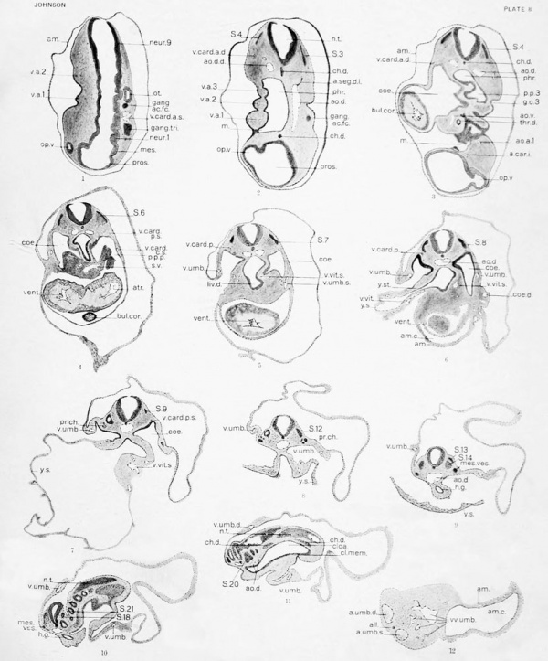
|
Abbreviations
a. cer., (?) a. car. i., a. seg. d., a. seg. I., a. aeg. v., a. umb.. a. uml). d., a. uuib. »., aa. uml)., all.,
am. c, ao. d., ao. d. (1., ao. d. !>., ao. v., ao. a., ao. p.,
a. pi., t., atr. p., atr., aud. p.,
h. c,
b. v., b. St.,
b. w., bul. cor.,
cap., cap. pi., r. int., ch. d., ch. can., ch. p., cloa. , d. men.,
c. m., coe.,
coe. cloa. d.,
coe. cloa. s.,
coe. d., coe. f. R., cop.,
d. b..
a. basilaris.
a. cerebri (?).
a. carotis interna.
a. .scgmentalis dorsalis.
a. segmentalis lateralis.
a. segmentalis ventralis.
a. umbilicalis.
a. umbilicalis dcxtra.
a. umbilicalis sinistra.
aa. umbilicalcs.
allantois.
amnion.
amnionic cavity.
aorta dorsalis.
aorta dorsalis dextrfl.
aorta dorsali.'; sinistra.
aorta ventralis.
aortic arch.
aortic process of sclerotome.
arterial plexus of tail.
atrial projection.
atrium.
auditory pit.
blood-corpuscles, blood-vessels, body-stalk, body-wall, bulbus cordis.
capillary.
capillary plexus.
caudal intestine.
chorda dorsalis.
chordal canal.
chordal process of sclerotome.
cloaca.
cloacal membrane.
cuticular membrane.
Cfrlom.
coplom on right side of cloaca and caudal
intestine, ccelom on left side of cloaca and caudal
intestine, ctrlomic depres.sions. ccplom surrounding forc-gul. copula.
dorsal branch of medial limb of a. seg. d.
ect..
ectoderm.
end. ht..
endothelial heart.
end. p..
endothelial process.
end..
endothelium.
ex. coe..
exoccelomic cavity.
f. g..
fore- gut.
gang. ac. fc,
ganglion acustico-facialis.
gang, c.
ganglion crest.
gang. gl. ph.,
ganglion n. glossopharj'ngei
gang, tri.,
ganglion n. trigemiui.
gang, va.,
ganglion n. vagi.
gas. r.,
gastric region.
g. c.
gill-cleft.
ht. sw..
heart swelling (Grosser).
h. g..
hind-gut.
inf., infundibulum.
int., integument.
i. e., intersegmental enlargement of v.
I. b., lateral branch of a. seg. d.
I. t., lateral tributary of v. card. p.
liv. d. (p. c), liver diverticulum (pars cystica),
liv. d. (p. h.), liver diverticulum (pars hepatica).
liv. tr., liver trabeculae.
Ig. d., lung diverticulum.
urd. p.
md. a.,
mandibular arch.
m. b..
medial branch of a. seg. d.
m. and 1. t..
medial and lateral tributaries of v. card, p
m. c,
mew)cardium.
mes..
mesencephalon.
mes. ves..
mesonephric vesicles.
meso.,
mcsothciium.
mit.,
mitotic figure.
m.,
mouth.
m. c..
myoepicardium.
myc..
myococle.
myt..
myotome.
nep.,
ncphrostome.
n. t..
medullary tube.
neur..
ncuromcre (1-21).
op. v.,
optic vesicle.
ot..
otocyst.
or. m..
oral membrane remnants.
p. d. mes..
phr., p. ex. d.,
p.
Ig. d., pr. ch., proc. inv., pros., p. V. mes., p. y. s., p. y. St.,
sclt..
t..
tear, thr. d., tr. a., t. imp., t. p. ex. d.,
u. aa. uml)., u. vv. umb.,
V. card. a. d.,
V. card. a. s.,
V. card. c. d.,
V. card. c. s.,
V. card. p. d.,
V. card. p. s.,
V. c. a., (?)
V. c. m.,
V. c. p..
V. ling-f..
V. oph.,
V. umb. d.,
V. umb. s..
V. vit. d.,
V. vit. s..
VV. a.,
vv. umb..
V. pi. b. w..
v. pi. h. g..
V. pi. y. St.,
V. b.,
V. p. gr..
V. p. gr. 1 (p. 1),
vent.,
vil..
pericardial cavity.
position of dorsal mesentery.
pleuro-pericardial passage.
pharyngeal pouch.
pharj'nx.
primary excretory- duct.
principal collecting tubule.
position of lung diverticulum.
proncphric chamber.
prortofleal invagination.
prosencephalon.
position of ventral mesentery.
position of yolk-sac.
position of yolk-stalk.
sclerotome, septum transversum. sinus sagit talis superior, sinus veno.sus. .somite (1-22).
tail.
tear in tissue.
thyroid diverticulum.
truficus arteriosus.
tuberculum impar.
termination of primary excretory duct.
union of aa. umbilicaleg. union of vv. umbilicalcs.
V. cardinalis anterior dextra.
V. cardinalis anterior sinistra.
V. cardinalis eonmiunis dcxtra.
V. cardinalis eomnumis sinistra.
V. cardinalis posterior dextra.
V. cardinalis posterior sinistra.
V. cerebri anterior (?).'
V. cerebri media.
V. cerebri posterior.
V. linguo-facialis.
V. ophthalamicus.
V. umbilicalis dextra.
V. umbilicalis sinistra.
V. vitellina dextra.
V. vitcUina sinistra.
veins of visceral arches (I-IV).
vena; umbilicalcs.
venous plexus of body-wall.
venous plexus of hind-gut.
venous plexus of yolk-stalk.
ventral branch of medial limb of a. seg. d.
ventral pharyngeal groove (1-3).
ventral pharyngeal groove 1-posteriorlimb.
ventricular portion of heart.
villus-like projections of body-wall.
visceral arch (1-3).
yolk-sac. yolk-stalk.
Bibliography
Hhaiiii.t. a , Iviti, Rprhcrrhes »ur 1>- vl^'vi-lu|)|KMncut du pancrtiis ct <iu foi*-. Jour, dc I'anat. et pliyiiiol, Paris, XXXI 1, 020-696, 3 pi.
Bremer, J. L., 1906. Description of a 4 mm. Imrimn oml)r>o. .\mer. Jour. Aiiat.. Bait., V. 459 480.
. 1914. The earliest blood-veiwels in man.
.Amer. Jour. .-Vnat., Phila., xvi, 447-17o.
BRou.t.v, I., 1S95. Beschreihung eines mcnsi'hiiclicn embryos von Ijeinahe 3 mm. LiuiKO mit spezieller BemorkunR ulicr die hei demselben l>ofindlichcn Hirnfallon. Morph. .\rb., Jena, v, 109 2(1.^), 2 pi.
Etehnop, .\.C. F., 1S99. Ily auncanalnotochordaldans rcnibr>on liumain. .\jiat. Anz., Jena, xvi, 131-14.J.
Evans, H. M., 1909. On tlie devcloi)ment of the aorUr, cardinal and umbilical vein.'*, and other blood-vessels of vertebrate embryos from capillaries. Anat. Record, Phila., iii, 498-518.
. 1909. On the earliest blood-vessels in the anterior limb buds of birds and their relation to the priinary subclavian artery. Amer. Jour. Anat., Phila., IX, 2S1-319, 9 pi.
. 1912. The development of the vascular system.
In Manual of Human Embryology (Keibel and Mall), Phila. and I,ond., ii, 570-709. Also, Handbuch d. Entwcklngsgesch. d. Menschen (Keibel & Mall), Leipzig. 1911, ii. 551-688.
Kei.ix, W., 1892. Zur Leber- und Pankreasentwicklung. Arch. f.-Anat. Entwcklngsgesch., Leipzig, 281-323.
, 1912. The development of the urinogenital organs. In Manual of Human Embryology, (Keibel and Mall), Phila. and Lond., ii, 752-979. AUo, Handl>uch d. Entwcklngsgesch. d. Menschen. (Keibel and Mall), Leipzig, 1911, ii, 732-955.
Frassi, L., 1908. Weitere Ergebnisse des Studiums eines jungen nienschlichen Eies in situ. Arch. f. mikr. Anat., Bonn, lxxi, 067-094.
Gage, S. P., 1906. A three weeks' human embryo with special reference to the brain and the nephric system. Amer. Jour. Anat., Bait., iv, 409-443, 5 pi.
Grafe, E., 1905. Beitrage zur Entwicklung dcr Urniere und ihrcr GcfjSssc beim Huhnchen, Arch. f. mikr. Anat., Bonn, i.xvn, 143-230, 5 pi.
Grosser, O., 1910. Development of the egg membranes and the placenta. In Manual of Human Embryology, (Keibel and Mall), Phila. and Lond., i, 91-179. Alto, Handbuch d. Entwcklngsgesch. d. Menschen (Keibel and Mall), Leipzig, 1910, i, 97-184.
. 1912. The development of the pharynx and organs of respiration. In Manual of Human Embryology (Keibel and Mall). Phila. and Lond., II, 446497. AUo, Handbuch d. Entwcklngsgesch. d. Menschen (Keibel and Mall), Leipzig, 1911, ii, 436-482.
. 1901. Zur Anatomic und Entwicklungsgeschichtc des Gefiisssystemes der Chiropteren. Anat. Hefle, 1. Abt., Wiesk, xvii, 203-124, 13 pi.
Hamuar, J. A., 1902. .Siudien i'lber die Entwicklung des Vonlcrdarms und einiger angrciizenilen Organe. An-h. f. mikr. Anat., Bonn, ux, 471-62K, 5 pi.
HiH, W., 18S5. Anatomic nienschlicher Embryonen, III, l^ipzig, F. f. W. Vogel.
Inoai.ls, N. W., 1907. Beschreibung eines mcDschlichen embryos von 4.9 mm. Arch. f. mikr. Anat., Bonn, i.xx, .IOfi-576, 3 pi.
Jan'ohik, J., 1887. Zwci jiuiKe mcnschlichc Embr}i'oncn. Arch. f. mikr. Anat., Bonn, xxx, .V>9 595, 2 pi.
JuiiMwiN, 1916. Notes on the neuronieres of brain anil spinal cord. Aiiut. Record. Vol. 10. Procco<liiig8 of the American' Association of Aiiatoniisls, pp. 209 210.
Kr.inRi., Fiu, 1910. Kumniary of the development of the human embryo and the difTerentintion of its external form. In Manual of Embryology (Keibel and Mall), Phila. aii<l Ixmd., I, .'>9-90. Al'ii, Hnndbuch d. Enlwcklngsgesch. d. Menschen (Keibel aii.l Mall), I*ip;
Kr.iiir.i., V. und ('. Eijie, KnlwickliinKKgewhlchte FiiM'her. . Ki>i.iJbiANN, J., IHOl. Die RumpfsCKnionte nieusch liehrr Kiiilirytmeii von 13 bis 35 nrwirlK-lii, Arch f. AiiBt. Rnlwrklnitigeitch. Lcipilg, .'19 88, 3 pi
n. I, «& 90. 1908. N'orinentiifel zur les Menschen, Jena, (i.
25. Lexhoksek. M. von. 1891. Die Entwicklung dcr Ganglienanlugen bei dem menschlichen Embryo. Arch. f. Anat. u. Entwcklngsgesch., Leipzig, 1-25,1 pi.
26. Lewis, F. T., 1903. The gross anatomy of a 12 mm. pig. .\mer. Jour. Anat., Bait., Ii, 211-225.
27. , 1909. On the cerWcal veins and lymphatics in four human embryos. Amer. Jour. Anat., Phila., IX, 33^2.
28. . 1912. The early development of the entodcrmal tract and the formation of its subdivisions. In Manual of Human Embryology, (Keibel and Mall), Phila. and Lond., ii, 29.5-3.34. Aho, Handbuch d. Entwcklngsgesch. d. Menschen (Keibel and Mall), Leipzig, 1911, ii, 286-324.
29. , 1912. The development of the liver. Ibid., II, 403-428 (.'igi-SlS respeclirely).
30. Mall, V. P., 1891. A human embryo twenty-six days old. Jour. Morphol., Bost., v, 459-480, 2 pi.
31. ■— ■, 1904. On the development of the blood-vessels of the brain in the human embryo. Amer. Jour. Anat., Bait., iv, 1-18, 3 pi.
32. , 1910. Determination of the age of human embryos and fetuses. In Manual_of Human Embryology (Keibel and Mall), Phila. and Lond., i, 180-201. Aho, Handbuch d. Entwcklngsgesch. d. Menschen (Keibel and Mall), Leipzig, i, 185-207.
33. , 1912. On the development of the human jheart. Amer. Jour. Anat., Phila., xiii. 249-298.
34. ., 1910. Ca-lom and [diaphragm, /n Manual of Human Embryology (Keibel and Mall), Phila. and Lond., I, 523-548. Also, Handbuch d. Entwcklngsgesch. d. Menschen (Keibel and Mall), Leipzig, I, 527-552.
35. Maurer, F., 1902. Die Entwicklung des Darmsystems. In: Handbuch d. vergleichenden u. experimentellen Entwicklungslehre d. Wirbeltiere (O. Hcrtwig), Jena, ii, 1. Teil, 109-252.
36. MiNOT, C. S., 1892. Human Embryology. New York, W. Wood & Co.
37. — ■— - — , 1907. The segmental flexures of the notochord. Anat. Record, Bait., i, 42-50.
38. Neal, J. v., 1914. The morphologj' of the eye-muscle nerves. Jour, of Morphology, xxv, pp. 1-187. 9 pis.
39. Phisalix, C, 1888. fitude d'un embrj-on humain de 10 millimitres. Arch, dc zool. exp6r. et g4n., s6r. 2, VI, 279-350.
40. Sai.zer, H., 1895. Ueber die Entwicklung der Kopfvenen des Meerschweinchcns, Morphol. Jahrb., Leipzig, xxiii, 232-255, 1 pi.
41. Smith, H. W., 1909. On the development of the superficial veins of the bodj'-wall in the pig. Amer. Jour. Anat., Phila., ix, 439-402.
42. Streeter, G. L., 1912. Development of the nervous system. In Manual of Human Embryology (Keibel and Mall), Phila. and Lond., ii, 1-156. AUo. Handbuch d. Entwcklngsgesch. d. Menschen (Keil>cl and NLall), Leipzig, 1911, ii, 1-156.
43. TANni.ER, J., 1907. Ueber einen nienschlichen Embryo vom 38. Tagc. Anat. Anz., Jena, xxxi, 49-56.
44. Tandler, J, 1912. The development of the heart. Manual of Human Embryology (Keibel and Mall), Phila. and I>ond., ii, 534-570. AUo, Handbuch d. Entwcklngsgesch. d. Menschen (Keibel and Mall), Leipzig, 1911, II, 517 551.
1.1. TiKi.MiwiN, Peter, 1907. Description of a Human Embryo of twenty-three paired somites. Jour. Anat. and PliJ'siol., Lond., xi.i, 1.59 171, 3 pi.
10. -, 1908. A note on the development of the Be|)lum Iransversum and the liver. Jour. Anat and Physiol., I/ond., XI.II, 170-175.
47. VAN den Broeck, a. J. P., 1911. Zur Kosuistik junger nienschlicher Embrj-onen. Anat. Hefte, 1. Abt., Wiesb., XLiv, 27.5-304, 5 pi.
48. Watt, J. <^, 1915. Description of two young twin human embryos with 17-19 paired somites. Contributions to Embryology No. 2, Carnegie Inst. Wash. Pub. No. 222.
49. WiiiiAMR, L. W., 1910. The somites of the chick. Amer. Jour. Anal., Phila., xi, ,55-100.
| Historic Disclaimer - information about historic embryology pages |
|---|
| Pages where the terms "Historic" (textbooks, papers, people, recommendations) appear on this site, and sections within pages where this disclaimer appears, indicate that the content and scientific understanding are specific to the time of publication. This means that while some scientific descriptions are still accurate, the terminology and interpretation of the developmental mechanisms reflect the understanding at the time of original publication and those of the preceding periods, these terms, interpretations and recommendations may not reflect our current scientific understanding. (More? Embryology History | Historic Embryology Papers) |
Glossary Links
- Glossary: A | B | C | D | E | F | G | H | I | J | K | L | M | N | O | P | Q | R | S | T | U | V | W | X | Y | Z | Numbers | Symbols | Term Link
Cite this page: Hill, M.A. (2024, April 28) Embryology Book - Contributions to Embryology Carnegie Institution No.19. Retrieved from https://embryology.med.unsw.edu.au/embryology/index.php/Book_-_Contributions_to_Embryology_Carnegie_Institution_No.19
- © Dr Mark Hill 2024, UNSW Embryology ISBN: 978 0 7334 2609 4 - UNSW CRICOS Provider Code No. 00098G

