User:Z3288196: Difference between revisions
No edit summary |
|||
| Line 70: | Line 70: | ||
--[[User:Z3288196|Z3288196]] 16:13, 17 August 2011 (EST) | --[[User:Z3288196|Z3288196]] 16:13, 17 August 2011 (EST) | ||
==Lab 4 Assessment== | |||
1. The allantois, identified in the placental cord, is continuous with what anatomical structure? | |||
The allantois is composed of an inner layer of endodermal cells and an outer layer of mesoderm. The inner layer is continuous with the endoderm of the digestive tract and the outer layer is continuous with the splanchnic mesoderm of the embryo. The allantois dilates with development into the allantoic sac and remains connected to the hindgut by the allantoic stalk, which passes through the umbilical cord. | |||
2. Identify the 3 vascular shunts, and their location, in the embryonic circulation. | |||
i) Ductus Arteriosus: Connects the pulmonary artery with the aortic arch. | |||
ii) Ductus Venosus: Connects the umbilical and portal veins to the inferior vena cava. | |||
iii) Foramen Ovale: Connects the right and left atria. | |||
3. Identify the Group project sub-section that you will be researching. (Add to project page and your individual assessment page) | |||
I am doing the Etiology and Further Research sub-sections in our group project on DiGeorge Syndrome. | |||
==Lab 5 Assessment== | |||
1. Which side (L/R) is most common for diaphragmatic hernia and why? | |||
The left side is most common for the occurrence of Congenital Diaphragmatic Hernia. Herniation refers to abnormal closure of the pleuroperitoneal foramen. When the pleuroperitoneal folds close the foramen, the right side usually closes first. Hence this increases the likelihood of an opening on the left side through which the viscera of the gut can move in to. | |||
--[[User:Z3288196|Timothy Ellwood]] 10:19, 1 September 2011 (EST) | |||
Revision as of 10:19, 1 September 2011
Lab 4 Online Assessment
- The allantois, identified in the placental cord, is continuous with what anatomical structure?
- Identify the 3 vascular shunts, and their location, in the embryonic circulation.
- Identify the Group project sub-section that you will be researching. (Add to project page and your individual assessment page)
--Z3288196 12:55, 28 July 2011 (EST)
--Mark Hill 09:38, 3 August 2011 (EST) All 3 questions from Lab 1 need to be completed before Lab 2.
Lab 1 Assessment
1. Identify the origin of In Vitro Fertilization and the 2010 nobel prize winner associated with this technique.
In Vitro Fertilization refers to the fertilization of an egg cell outside of the organism, usually in a laboratory environment. Robert G. Edwards was the first to successfully perform this process in 1978, and was awarded the Nobel Prize in Physiology or Medicine in 2010 as recognition for his work in developing this method of fertilization.
2. Identify a recent paper on fertilisation and describe its key findings.
Groeneveld, E.; Broeze, K. A.; Lambers, M. J.; Haapsamo, M.; Dirckx, K.; Schoot, B. C.; Salle, B.; Duvan, C. I. et al. (2011). "Is aspirin effective in women undergoing in vitro fertilization (IVF)? Results from an individual patient data meta-analysis (IPD MA)". Human Reproduction Update 17 (4): 501–509.
The key finding of this paper is that aspirin is not effective in women undergoing IVF. Women who take aspirin after IVF did not show improved pregnancy rates after IVF.
3. Identify 2 congenital anomalies.
- Congenital Diaphragmatic Hernia
- Spina Bifida
--Z3288196 09:23, 4 August 2011 (EST)
Lab 2 Assessment
1. Identify the ZP protein that spermatozoa binds and how is this changed (altered) after fertilization.
Spermatozoa bind to Zona Pellucida Glycoprotein 3 (ZP3). After fertilization has occured, enzymes digest the zona pellucida and spermatozoa are no longer able to bind to ZP3 as it is altered by these digestive enzymes.
2. Identify a review and a research article related to your group topic. (Paste on both group discussion page with signature and on your own page)
The primary journal article I found talks about the expression of the DMD Gene Products in Embryonic Stem Cells, so I'm hoping that this will allow our group to elaborate a bit on the genetic aspects of the development and potential early identification, as symptoms do not usually appear in humans until a few years of age [1]. Also the review article I found refers to two other types of muscular dystrophies as well as DMD (SCARMD, CMD), highlighting many key developments in research. Seem to be very informative, also refers a bit to the importance of animal models in these developments [2].
--Z3288196 12:38, 4 August 2011 (EST)
Lab 3 Assessment
1.What is the maternal dietary requirement for late neural development?
NHRMC Policy (1993) suggests that an intake of 0.4mg-0.5mg of folate is required to reduce incidence of neural tube defects, such as spina bifida and anencephaly, and allow for normal neural development.
--Z3288196 12:35, 11 August 2011 (EST)
2. Upload and post an image.
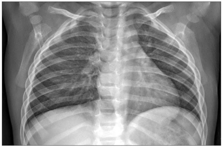
"Aspiration pneumonia in the child with DiGeorge syndrome -- A case report "
A preoperative chest PA showing a narrowed superior mediastinum suggesting thymic agenesis, apical herniation of the right lung and a resultant left sided buckling of the adjacent trachea air column.
Ji-Young Lee, Yun-Joung Han "Aspiration pneumonia in the child with DiGeorge syndrome -A case report-"
Korean J Anesthesiol. 2011 June; 60(6): 449–452. Published online 2011 June 17. doi: 10.4097/kjae.2011.60.6.449 [3]
This is an open-access article distributed under the terms of the Creative Commons Attribution Non-Commercial License [4], which permits unrestricted non-commercial use, distribution, and reproduction in any medium, provided the original work is properly cited.
--Z3288196 16:13, 17 August 2011 (EST)
Lab 4 Assessment
1. The allantois, identified in the placental cord, is continuous with what anatomical structure?
The allantois is composed of an inner layer of endodermal cells and an outer layer of mesoderm. The inner layer is continuous with the endoderm of the digestive tract and the outer layer is continuous with the splanchnic mesoderm of the embryo. The allantois dilates with development into the allantoic sac and remains connected to the hindgut by the allantoic stalk, which passes through the umbilical cord.
2. Identify the 3 vascular shunts, and their location, in the embryonic circulation.
i) Ductus Arteriosus: Connects the pulmonary artery with the aortic arch. ii) Ductus Venosus: Connects the umbilical and portal veins to the inferior vena cava. iii) Foramen Ovale: Connects the right and left atria.
3. Identify the Group project sub-section that you will be researching. (Add to project page and your individual assessment page)
I am doing the Etiology and Further Research sub-sections in our group project on DiGeorge Syndrome.
Lab 5 Assessment
1. Which side (L/R) is most common for diaphragmatic hernia and why?
The left side is most common for the occurrence of Congenital Diaphragmatic Hernia. Herniation refers to abnormal closure of the pleuroperitoneal foramen. When the pleuroperitoneal folds close the foramen, the right side usually closes first. Hence this increases the likelihood of an opening on the left side through which the viscera of the gut can move in to.
--Timothy Ellwood 10:19, 1 September 2011 (EST)

