Ultrasound - Fetus Movie 2: Difference between revisions
mNo edit summary |
mNo edit summary |
||
| Line 2: | Line 2: | ||
{| border='0px' | {| border='0px' | ||
|- | |- | ||
| <mediaplayer width=' | | <mediaplayer width='400 height='530' image="http://embryology.med.unsw.edu.au/embryology/images/2/25/Ultrasound12wk_3D_image2.jpg">File:Ultrasound_Fetus_02.mp4</mediaplayer> | ||
| valign="top" | | | valign="top" | | ||
===Fetus (12 weeks)=== | ===Fetus (12 weeks)=== | ||
Revision as of 16:08, 16 March 2013
| Embryology - 4 May 2024 |
|---|
| Google Translate - select your language from the list shown below (this will open a new external page) |
|
العربية | català | 中文 | 中國傳統的 | français | Deutsche | עִברִית | हिंदी | bahasa Indonesia | italiano | 日本語 | 한국어 | မြန်မာ | Pilipino | Polskie | português | ਪੰਜਾਬੀ ਦੇ | Română | русский | Español | Swahili | Svensk | ไทย | Türkçe | اردو | ייִדיש | Tiếng Việt These external translations are automated and may not be accurate. (More? About Translations) |
| <mediaplayer width='400 height='530' image="http://embryology.med.unsw.edu.au/embryology/images/2/25/Ultrasound12wk_3D_image2.jpg">File:Ultrasound_Fetus_02.mp4</mediaplayer> |
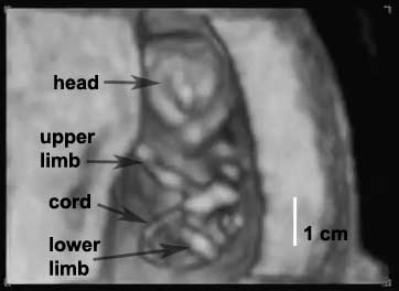
Fetus (12 weeks)Movie shows a 12 week fetus in 3d in realtime (hence 4D). The beginning of the movies shows the ventral (anterior) view of the fetus head to top.
Arrow indicates: initially head, then upper limbs, then lower limbs, then umbilical cord and finally the placenta.
The second part of movie shows fetal movements. Calibration visible on left shows 1 cm increments.
|
Glossary Links: A | B | C | D | E | F | G | H | I | J | K | L | M | N | O | P | Q | R | S | T | U | V | W | X | Y | Z | Numbers | Symbols | Movies
Cite this page: Hill, M.A. (2024, May 4) Embryology Ultrasound - Fetus Movie 2. Retrieved from https://embryology.med.unsw.edu.au/embryology/index.php/Ultrasound_-_Fetus_Movie_2
- © Dr Mark Hill 2024, UNSW Embryology ISBN: 978 0 7334 2609 4 - UNSW CRICOS Provider Code No. 00098G