Template:2012GroupDiscussion
Assessment
Images
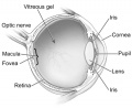
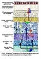
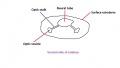
Basic structure of the human eye.

Basic anatomy of the eye.
![Illustration of the front of the eye, showing the iris, sclera and pupil. Credits: Webvision PMID:21413389 [PubMed]](/embryology/images/thumb/c/c4/Eye-pupil-sclera-iris.jpg/120px-Eye-pupil-sclera-iris.jpg)
Illustration of the front of the eye, showing the iris, sclera and pupil. Credits: Webvision PMID:21413389 [PubMed]
![A CAT scan with illustrations to show the extraocular muscles from the back view of the eye. Credits: Webvision PMID:21413389 [PubMed]](/embryology/images/thumb/9/93/Extraocular-muscles-scan.jpg/120px-Extraocular-muscles-scan.jpg)
A CAT scan with illustrations to show the extraocular muscles from the back view of the eye. Credits: Webvision PMID:21413389 [PubMed]
![A diagram of the layers of the retina. Credits: Webvision PMID:21413389 [PubMed]](/embryology/images/thumb/d/d6/Retina-layers-diagram2.jpg/120px-Retina-layers-diagram2.jpg)
A diagram of the layers of the retina. Credits: Webvision PMID:21413389 [PubMed]
![The layers of the retina magnified, showing the direction of the layers of the retina in the back of the eye. Credits: Webvision PMID:21413389 [PubMed]](/embryology/images/thumb/8/8f/Eye-retina-layers.jpg/120px-Eye-retina-layers.jpg)
The layers of the retina magnified, showing the direction of the layers of the retina in the back of the eye. Credits: Webvision PMID:21413389 [PubMed]
![A diagram of the components of the retina. Credits: Webvision PMID:21413389 [PubMed]](/embryology/images/thumb/5/57/Retina-layers-diagram.jpg/120px-Retina-layers-diagram.jpg)
A diagram of the components of the retina. Credits: Webvision PMID:21413389 [PubMed]
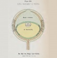
The eye according to Aristotle. Credits: Magnus, 1901. Note the lens is missing, and there are three vessels drawn that was believed to transport fluid to and from the eye.
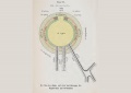
The eye according to Celsus. Credits: Magnus, 1901. Note the lens is placed in the centre of the eye, in the vitreous.
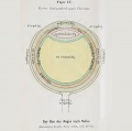
The eye according to Rufus of Ephesus. Credits: Magnus, 1901. Note the lens is placed in the correct position, behind the iris of the eye
The eye according to Galen. Credits: Magnus, 1901.
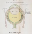
The blue part at the bottom is the endoderm. The pink middle layer is the mesoderm. The top yellow layer is the ectoderm. The fold labelled as 'augenfeld' is the place where the optic vesicle will form. Credits: Kollmann, J. (1907)
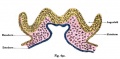
The eye area (augenfeld) is a bowl shaped bulge still located on the side walls. Credits: Kollmann, J. (1907)
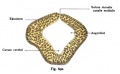
The neural tube is shown after removal of all of the ectoderm and ventral organs, such as heart, gut tube, etc. The primary optic vesicle forms a slightly flattened hollow protrusion on the forebrain. Credits: Kollmann, J. (1907)
The lateral surface of the primary optic vesicle is slightly depressed, showing the first sign of the emergence of the secondary optic vesicle. Credits: Kollmann, J. (1907)
The bulging lateral wall of the primary optic vesicle is covered by a fairly well demarcated lens plate, a direct continuation of the ectoderm. Between the optic vesicle and the lens pit are some flattened spindle-shaped cells. In the adjoining mesoderm are cross-sections of capillaries. Credits: Kollmann, J. (1907)
The lens still hangs together with the ectoderm. The primary eye vesicle is indented with respect to the lens. Between the lens and the lateral plate of the optic vesicle is a narrow space, which allows area to further develop later. Credits: Kollmann, J. (1907)

4th Week of development. The internal organisation shows the secondary optic vesicle. A: The rear wall of lens is noticeable and is enveloped by mesoderm. B: The edges of the lens pit is already grown and the lens vesicles are formed, which is still related to the remaining ectoderm. Credits: Kollmann, J. (1907)
The lens has now cut off from the ectoderm, but is still very superficial. Between it and the lateral lamina of the optic cup, there is a considerable space. The eye stalk has become longer and is enclosed together with the optic cup and lens of the mesoderm. The cornea, sclera and choroid make gradual development. Credits: Kollmann, J. (1907)
The layers of the retina in the fifth month of development. Credits: Webvision PMID:21413389 [PubMed
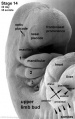
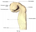
A Stage 14 embryo showing the location of an otic placode.
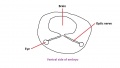
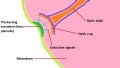
A cross section showing the organisation of the developing brain, the optic vesicle and the lens (optic) placode.
Early formation of the optic vesicle from the neural groove.
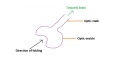
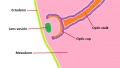
The optic vesicle at a later stage, showing the optic stalk.
A recognisable brain and eye structure in later development.
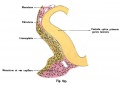
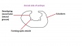
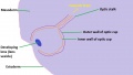
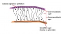
Mechanism of optic cup formation.
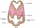
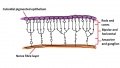
Layers of the optic cup in retina development.
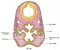
Cross-section of the primitive retina showing cell types and layers.
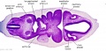
Cross-section of a developed retina showing cell types and layers.
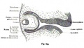
The importance of the optic cup in lens differentiation.
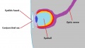
The lens placode separates from the ectoderm and migrates into the mesoderm forming the lens vesicle.
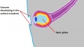
The choroid and sclera derives from mesenchyme surrounding the optic cup.
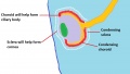
Small grooves in the ectoderm of the head - the precursors to an eyelid.
The eye at an advanced stage of embryonic development. Note however, that the eyelids remain fused until much later.
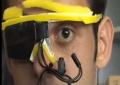
An early prototype of the bionic eye.


![Illustration of the front of the eye, showing the iris, sclera and pupil. Credits: Webvision PMID:21413389 [PubMed]](/embryology/index.php?title=File:Eye-pupil-sclera-iris.jpg)
![A CAT scan with illustrations to show the extraocular muscles from the back view of the eye. Credits: Webvision PMID:21413389 [PubMed]](/embryology/index.php?title=File:Extraocular-muscles-scan.jpg)
![A diagram of the layers of the retina. Credits: Webvision PMID:21413389 [PubMed]](/embryology/index.php?title=File:Retina-layers-diagram2.jpg)
![The layers of the retina magnified, showing the direction of the layers of the retina in the back of the eye. Credits: Webvision PMID:21413389 [PubMed]](/embryology/index.php?title=File:Eye-retina-layers.jpg)
![A diagram of the components of the retina. Credits: Webvision PMID:21413389 [PubMed]](/embryology/index.php?title=File:Retina-layers-diagram.jpg)


From the Course Coordinator
- 2012 Projects: Vision | Somatosensory | Taste | Olfaction | Abnormal Vision | Hearing
--Mark Hill 09:58, 18 September 2012 (EST) This is a recent review on vision. http://jcb.rupress.org/content/190/6/953.full JCB content allows reuse.
http://www.jove.com/video/3730/isolation-and-culture-of-human-fungiform-taste-papillae-cells
--Mark Hill 08:17, 16 August 2012 (EST) This small section at the top of your discussion page allows me to easily distribute information to all students looking at the group discussion page.
- Please do not delete, edit or move the template {{Template:2012GroupDiscussion}} from the top of this page.
1. Search:
- http://www.ncbi.nlm.nih.gov/sites/gquery?term=embryo Pubmed search all databases term embryo, replace this with your own term to set up a search on your topic.
- http://www.ncbi.nlm.nih.gov/pmc/?term=embryo&report=imagesdocsum Pubed Central search for images using the term embryo, replace this with your own term to set up a search on your topic.
2. History
- Embryology History Explore the rich history of sensory research. In particular look through the available images that may complement the text. Any of this material can be used, you should cite the original paper or textbook. Gray | 1921 Bailey and Miller