Talk:SH Lecture - Lymphatic Structure and Organs
SH Laboratory - Lymphatic Structure and Organs
Introduction
This practical class has 2 main parts.
The first part will look at the cellular components of blood and their development.
The second part will look at the organs and tissues associated with lymphoid (lymphatic) immune function.
The Practical involves studying selected slides from the Virtual Slide Box (some text in these practical notes modified from Virtual Slidebox Histology). Additional online self-directed learning resources are shown below. Today's Laboratory will not cover all the immune system associated with the gastrointestinal tract (GIT), just the tonsilal component.
Objectives
1. Understand the major immune cell types of blood as they appear in blood smears
2. Brief understanding of the appearance and function of blood cells
3. Understand the organization of lymphoid tissue
4. Brief understanding of the organization of thymus, spleen and tonsil
Online References
- Dee, F.R., Leaven, T. and Consoer, D. (2005) Virtual Slidebox of Histology - Hematopoietic system. Retrieved April 14, 2005, from http://www.path.uiowa.edu/cgi-bin-pub/vs/fpx_browse.cgi?cat=o_hemato&div=nlm
- UWA Blue Histology UWA Blue Histology | Blood | Lymphoid Tissues 1 | Lymphoid Tissues 2 | | appendix
UNSW Virtual Slidebox
- http://vslide1.med.unsw.edu.au/B-blood-lymphoid.html or http://web.med.unsw.edu.au/vslide/phase1/indexB3.asp
- This new link does not function http://vslide.med.unsw.edu.au/index.jsp
Blood Cell Data
Bone Marrow
Promyelocyte -> Myelocyte -> Metamyelocyte -> White Blood Cell type
Neutrophil "band form" stage before final maturation
Online Resources
UWA Blue Histology UWA Blue Histology | Blood | Lymphoid Tissues 1 | Lymphoid Tissues 2
National Institute of Allergy and Infectious Diseases National Institute of Allergy and Infectious Diseases - Immune System | Organs of the Immune System | Lymph Node | Lymphatic Vessel | Understanding the Immune System: How It Works (PDF)
JayDoc Web lymphoid
SEER Training Modules
Anatomy of the Human Body. 1918
Henry Gray (1821–1865).
THE LYMPHATIC SYSTEM consists (1) of complex capillary networks which collect the lymph in the various organs and tissues; (2) of an elaborate system of collecting vessels which conduct the lymph from the capillaries to the large veins of the neck at the junction of the internal jugular and subclavian veins, where the lymph is poured into the blood stream; and (3) lymph glands or nodes which are interspaced in the pathways of the collecting vessels filtering the lymph as it passes through them and contributing lymphocytes to it. The lymphatic capillaries and collecting vessels are lined throughout by a continuous layer of endothelial cells, forming thus a closed system. The lymphatic vessels of the small intestine receive the special designation of lacteals or chyliferous vessels; they differ in no respect from the lymphatic vessels generally excepting that during the process of digestion they contain a milk-white fluid, the chyle.
Lymphatic Vessels
The lymphatic vessels are exceedingly delicate, and their coats are so transparent that the fluid they contain is readily seen through them. They are interrupted at intervals by constrictions, which give them a knotted or beaded appearance; these constrictions correspond to the situations of valves in their interior. Lymphatic vessels have been found in nearly every texture and organ of the body which contains bloodvessels. Such non-vascular structures as cartilage, the nails, cuticle, and hair have none, but with these exceptions it is probable that eventually all parts will be found to be permeated by these vessels.
Lymph Glands
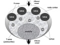
The lymph glands are small oval or bean-shaped bodies, situated in the course of lymphatic and lacteal vessels so that the lymph and chyle pass through them on their way to the blood. Each generally presents on one side a slight depression—the hilus—through which the bloodvessels enter and leave the interior. The efferent lymphatic vessel also emerges from the gland at this spot, while the afferent vessels enter the organ at different parts of the periphery. On section (Fig. 597) a lymph gland displays two different structures: an external, of lighter color—the cortical; and an internal, darker—the medullary. The cortical structure does not form a complete investment, but is deficient at the hilus, where the medullary portion reaches the surface of the gland; so that the efferent vessel is derived directly from the medullary structures, while the afferent vessels empty themselves into the cortical substance.
Lymph
Lymph, found only in the closed lymphatic vessels, is a transparent, colorless, or slightly yellow, watery fluid of specific gravity about 1.015; it closely resembles the blood plasma, but is more dilute. When it is examined under the microscope, leucocytes of the lymphocyte class are found floating in the transparent fluid; they are always increased in number after the passage of the lymph through lymphoid tissue, as in lymph glands. Lymph should be distinguished from “tissue fluid” 109 which is found outside the lymphatic vessels in the tissue spaces.
Spleen
http://www.bartleby.com/107/278.html
The splenic pulp (pulpa lienis) is a soft mass of a dark reddish-brown color, resembling grumous blood; it consists of a fine reticulum of fibers, continuous with those of the trabeculæ, to which are applied flat, branching cells. The meshes of the reticulum are filled with blood, in which, however, the white corpuscles are found to be in larger proportion than they are in ordinary blood. Large rounded cells, termed splenic cells, are also seen; these are capable of ameboid movement, and often contain pigment and red-blood corpuscles in their interior. The cells of the reticulum each possess a round or oval nucleus, and like the splenic cells, they may contain pigment granules in their cytoplasm; they do not stain deeply with carmine, and in this respect differ from the cells of the Malpighian bodies. In the young spleen, giant cells may also be found, each containing numerous nuclei or one compound nucleus. Nucleated red-blood corpuscles have also been found in the spleen of young animals.
Original Page

Introduction


--Mark Hill 11:30, 22 December 2010 (EST) This lecture is not complete and still under development for a 2011 course. The lecture will introduce the anatomical and cellular components of the immune system. The lecture will not cover development of the immune system or details on specific immune cell function.
- SH Links: Lymphatic Lecture | Lymphatics Practical Support | Respiratory Lecture | Respiratory Practical Support | Medicine
Start Time/End Time: 10am to 11am Monday 28 February 2011 Clancy Auditorium eMed Link to Learning Activity - Lymphatic organs histology
Aim
This lecture will provide an overview of the histology of key lymphoid organs, including the lymph nodes, spleen and thymus, as well as extranodal lymphoid tissues including mucosal associated lymphoid tissues (MALT)
Key Concepts
- Lymphatic System
- Organs - Thymus, Spleen
- Lymph Nodes and Nodules
- Bone Marrow
- Extranodal Lymphoid Tissues
- Mucosal Associated Lymphoid Tissues (MALT)
Textbook References
- Histology and Cell Biology - A.L. Kiersenbaum (2001)
- Chapter 6: Blood
- Chapter 10: Immune-Lymphatic
- Janeway’s Immunobiology NCBI Bookshelf | Publisher page
- SH Laboratory this week
- Online - Lecture 2010 | Online - Lecture 2008
Two Systems
- Lymphoid System - three major types of lymphocytes (T, B, and NK), tissues, organs and vessels
- Mononuclear Phagocytic System (MPS, also called Lymphoreticular System or Reticuloendothelial System, RES) - circulating monocytes of peripheral blood and non-circulating (fixed) tissue macrophages found throughout the body
MBoC Figure 24-3 Human lymphoid organs
Lymphatic System
- Connective Tissue Embryonic origin- Mesoderm
- Consists of Cells, tissues and organs
- Immune “monitor” of body surfaces, internal fluids
Immune System Note: Immunity is covered in detail elsewhere in the course current lecture is about Lymphoid Organ structure/location
- Tissues and Organs
- Thymus, spleen, lymph nodes, lymphatic nodules, diffuse lymphatic tissues, bone marrow
- Organs consist also of structural cells and extracellular matrix
- Lymphatic vessels connect system parts
- Cells are Lymphocytes
- B Lymphocytes and T Lymphocytes MBoC Figure 24-7. Electron micrographs of nonactivated and activated lymphocytes
- White blood cells, leukocytes
These are blood cells
Blood Cells


 large granular lymphocytes - natural killer (Nk-) cells
Central/Peripheral Lymphoid Organs
Central lymphoid organs
- Lymphocytes develop from precursor cells
Peripheral lymphoid organs
- Lymphocytes respond to antigen
- lymph nodes or spleen
Mononuclear Phagocytic System
(Mononuclear Phagocytic System MPS, also called Lymphoreticular System or Reticuloendothelial System, RES)


Mononuclear Phagocytes 2 types:
- Circulating monocytes of peripheral blood (monocytes entering the connective tissue differentiate into macrophages)
- Non-circulating (fixed) tissue macrophages found throughout the body (Liver (Kuffler cells), spleen and other tissues)
Lymph
- Fluid portion of lymphatic circulation
- blood plasma will leave blood vessels into surrounding tissues
- adds to normal tissue interstitial fluid
- surplus of liquid needs to be returned to circulation
- Lymph vessels are provide unidirectional flow of this liquid
Lymph Vessels


Three types based on size and morphology
- Lymph capillaries begin as blind-ending tubes in connective tissue, larger than blood capillaries, very irregularly shaped
- Lymph collecting vessels larger and form valves, morphology similar to lymph capillaries
- Lymph ducts 1 or 2 layers of smooth muscle cells in wall
(Remember anatomy NAVL = Nerve, Artery, Vein and Lymph)
Lymphocyte Circulation
- The circulation through a lymph node is shown.
- Microbial antigens are carried into the lymph node by dendritic cells, which enter via afferent lymphatic vessels draining an infected tissue.
- T and B cells, by contrast, enter the lymph node via an artery and migrate out of the bloodstream through postcapillary venules.
- Unless they encounter their antigen, the T and B cells leave the lymph node via efferent lymphatic vessels, which eventually join the thoracic duct.
- The thoracic duct empties into a large vein carrying blood to the heart.
- A typical circulation cycle takes about 12–24 hours.
Links: MBoC Chapter 24 - The Adaptive Immune System | MBoC Figure 24-14. The path followed by lymphocytes as they continuously circulate between the lymph and blood | Immunobiology
Diffuse Lymphatic Tissue
Alimentary canal, respiratory passage, urogenital tract
- Not enclosed by a capsule
- Located in subepithelial tissue - Lamina propria
Lymphocytes
- travel to nodes and back again
- proliferation and differentiation
Effector cells
- B Cell secreting antibody = Plasma Cell
- T Cell = Memory Cell
- Diffuse lymphatic tissue + nodules
- Reactive - enlarge when activated (by antigen)
MALT, BALT and GALT

Internal epithelia Associated Lymphoid Tissue - naming based upon the anatomical locations
- MALT - Mucosa Associated Lymphoid Tissue
- BALT - Bronchus Associated Lymphoid Tissue
- GALT - Gut Associated Lymphatic Tissue
Immune Responses
Adaptive immunity has 2 main classes
- Antibody-mediated - B Lymphocyte
- Cell-mediated - T Lymphocyte
Lymph Nodules
(or follicles)
- Organized concentrations of Lymphocytes
- No capsule, covered by epithelia
- Nodules are also unit structure seen in a node
- Oval concentrations in meshwork of reticular cells
Reticular cell
- produces reticular fibers (collagen type III) and surrounds the fibers with its cytoplasm
- reticular fibers are also produced by fibroblasts
Gastrointestinal Tract
- Oropharynx - Tonsils
- Distal small intestine (ilieum) - Peyer’s Patches
Appendix, cecum
Nodule States
- Primary Nodule - Mainly small lymphocytes
- Secondary Nodule
- Central pale region (germinal centre) - Effector cells and macrophages
- Dark outer ring (small lymphocytes)
Links: Immnuobiology - Figure 1.10. Organization of typical gut-associated lymphoid tissue
Tonsils
Anatomical location - Palatine (tonsils), Lingual and Pharyngeal ( adenoids )
Palatine Tonsils
File:Tonsil 01.jpg [[File:Tonsil_02.jpg]
- the "tonsils", lateral wall of oropharynx
- covered by stratified squamous epithelium
- numerous crypts (10-20) infolds of surface epithelium
- Afferent lymph vessels absent
- Efferent lymph vessels are present
Lingual Tonsils
- lamina propria root of tongue
- covered by stratified squamous epithelium
- salivary glands and skeletal muscle are directly adjacent
Pharyngeal Tonsils
- adenoids or nasopharyngeal tonsils, upper posterior part of throat
- covered by a pseudostratified ciliated epithelium with goblet cells
Peyer's Patch


Peyer's Patch, Ileum
microfold cells or M-cells
Lymph Nodes


Immunobiology - Figure 1.8. Organization of a lymph node
MBoC Figure 24-16. A simplified drawing of a human lymph node
- Encapsulated organ (1 mm - 2 cm)
- In lymph vessel pathways “filter”
- Afferent- towards node
- Efferent- away from node
- Location throughout the entire body - Concentrated in axilla, groin, mesenteries
- Antigen transformed lymphocytes from the blood
Lymph Node Structure
- Capsule - dense connective tissue
- Trabeculae - dense connective tissue
- Reticular Tissue - Reticular cells and fibers, supporting meshwork
File:Lyn041he.jpg File:Lyn10he.jpg
Lymph
- enters the node through afferent vessels
- filters through the sinuses
- leaves through efferent vessels
Subcapsular sinus = marginal sinus
Continuation of trabecular sinus
Lymph Node Development
Lymph Node Vessels
Lymphocyte Traffic
Links: Immunobiology - Figure 1.8. Organization of a lymph node | Blue Histology - Lymph Nodes
Thymus

MBoC Figure 24-6. The development and activation of T and B cells
Figure 24-7. Electron micrographs of nonactivated and activated lymphocytes
Development Changes


Changes with age Overall Size
- birth 10-15 g
- puberty 30-40 g
- after puberty - involution
- Replaced by adipose tissue
- middle-aged 10 g
Thymus Anatomy
- Superior mediastinum, anterior to heart
- Bilobed lymphoepithelial organ
- Contains reticular cells but no fibers
- Stem lymphocytes
- proliferate and differentiate
- forms long-lived T- lymphocytes
Thymus Cells
- Reticular cells
- Abundant, eosinophilic, large, ovoid and light nucleus 1-2 nucleoli
- sheathe cortical capillaries
- form an epitheloid layer
- maintain microenvironment for development of T-lymphocytes in cortex (thymic epitheliocytes)
- Macrophages
- cortex and medulla
- difficult to distinguish from reticular cells in H&E
- Lymphocytes
- cortex and medulla - more numerous (denser) in cortex
- majority of them developing T-lymphocytes (= thymic lymphocytes or thymocytes)
Fetal/Young Thymus
Thymic corpuscle
Hassall’s corpuscle - Mass of concentric epithelioreticular cells
Adult Thymus
Cortical lymphoid tissue replaced by adipose tissue Increase in size of thymic corpuscles
Links: Blue Histology - Thymus
Spleen
- Like- lymph nodes (filter)
- Unlike- is in blood stream
Structure
- Capsule, trabeculae (dense connective tissue)
- Splenic pulp White pulp, red pulp Based on appearance and cell content
Splenic Pulp
File:Spl041he.jpg File:Spl042he.jpg
White Pulp
- lymphocytes surround central arteries
- as periarterial lymphoid sheath (PALS)
Red Pulp
- Red blood cells
- Splenic sinuses
- Splenic cords
Spleen Structure
Reticular Fibers
File:Spl10re.jpg File:Spl43re.jpg
Spleen Reticular Fibres
Links: Immunobiology - Figure 1.9. Organization of the lymphoid tissues of the spleen
B Cell Development
- Bone marrow
- blood
- Lymph node, nodule
- Lymphatic vessel
- Bone marrow
Germinal Centres
- Bone Marrow
- Medullary cords contain plasma cells
Plasma cells
- secrete antibody directly into blood for distribution to all body
- in local extrafollicular sites are short lived 2–4 days
- longer-lived plasma cells in bone marrow 3 weeks to 3 months+
Additional Information
The following is not part of the lecture and is for reference purposes only.
- Intro to Associated Practical
- Blood Cells
- Florence R. Sabin (1871-1953)
Blood Cell Numbers
Red Blood Cells
- Male: 4.32 - 5.66 x 10¹²/l
- Female: 3.88 - 4.99 x 10¹²/l
Leukocytes
- Male: 3.7 - 9.5 x 10⁹/l
- Female: 3.9 - 11.1 x 10⁹/l
Granulocytes (1.8 - 8.9 x 10⁹/l)
- Neutrophils: 1.5 - 7.4 x 10⁹/l
- Eosinophils: 0.02 - 0.67 x 10⁹/l
- Basophils: 0 - 0.13 x 10⁹/l
Lymphocytes (1.1 - 3.5 x 10⁹/l)
- B-cells: 0.06 - 0.66 x 10⁹/l
- T-cells: 0.77 - 2.68 x 10⁹/l
- CD4+: 0.53 - 1.76 x 10⁹/l
- CD8+: 0.30 - 1.03 x 10⁹/l
- NK cells: 0.20 - 0.40 x 10⁹/l
Monocytes (0.21 - 0.92 x 10⁹/l)
Platelets
- 140 - 440 x 109/l
Textbook
Immunobiology by Janeway, Charles A.; Travers, Paul; Walport, Mark; Shlomchik, Mark New York and London: Garland Science; c2001
- Immunobiology
- Figure 1.7. The distribution of lymphoid tissues in the body
- Figure 10.18. Anatomy of mucosal immune responses
Anatomy of the Human Body (Gray)1918
- Historic anatomy is good, there are there are some functional inaccuracies.
- The Lymphatic System | The Thoracic Duct | The Spleen | The Thymus |
Lymph Node Analysis
- 3D Reconstruction Pelvic Node Anatomy
- Reconstruction of Axillary Node Anatomy
Additional Images
Lymph node cartoon
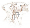
Lymph nodes - head neck superficial
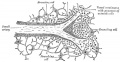
Section of the spleen, showing the termination of the small blood vessels


Terms
A few key terms associated with the Lymphoid system.
- adenoid - (Greek " +-oeides = in form of) in the form of a gland, glandular; the pharyngeal tonsil.
- Afferent lymph - vessel carrying lymph towards a node.
- Antibody mediated immunity - the immune function of plasma cells (active B lymphocytes) secreting antibody which binds antigen.
- antibodies - mammals have five classes (IgA, IgD, IgE, IgG, and IgM)
- antigen - any substance that is recognised by the immune system and stimulates antibody production.
- B lymphocyte (cell) - historically named after a structure called the bursa of Fabricius in birds, a source of antibody-producing lymphocytes. These cells develop in the bone marrow. (More? Electron micrographs of nonactivate and activated lymphocytes)
- BALT - Bronchus Associated Lymphoid Tissue
- band cell - (band neutrophil or stab cell) seen in bone marrow smear, a cell undergoing granulopoiesis, derived from a metamyelocyte, and leading to a mature granulocyte. Also occasionally seen in circulating blood.
- cell - has a specific cell biology definition, but is often used instead of "lymphocyte" when describing B and T cells.
- Cell-mediated immunity - the immune function of T lymphocytes.
- "clockface" - a term used to describe the appearance of plasma cell nuclei due to the clumping of the chromatin at the nucleus periphery. More clearly seen in tissue plasma cells that the bone marrow smear, where they are sometimes confused with the basophilic erythroblasts.
- cords of Billroth - spleen cellular columns located in red pulp. surrounded by splenic sinusoids. Cords contain reticular cells, macrophages, lymphocytes, plasma cells and erythrocytes.
- cortex - outer layer, used in association with medulla (innner layer or core) a general description that can be applied to describing an organ with a layered structure.
- Effector cells - the immune functioning (active) B and T lymphocytes.
- Efferent lymph - vessel carrying lymph away from a node.
- GALT - Gut Associated Lymphatic Tissue
- haemopoiesis (hemopoiesis) formation of blood cells.
- Hassall's corpuscle - thymic corpuscle.