Talk:Advanced - Outflow Tract: Difference between revisions
(Created page with '==Background Reading== * Septation and separation within the outflow tract of the developing heart Sandra Webb, Sonia R Qayyum, Robert H Anderson, Wouter H Lamers, and Michael K…') |
No edit summary |
||
| (10 intermediate revisions by 3 users not shown) | |||
| Line 1: | Line 1: | ||
{{Movie header}} | |||
<Flowplayer height="564" width="720" autoplay="false">outflow_tract_001.flv</Flowplayer> | |||
--[[User:S8600021|Mark Hill]] 18:09, 7 October 2009 (EST) [[File:Outflow_Tract_A.ppt]] | |||
--[[User:S8600021|Mark Hill]] 15:56, 2 October 2009 (EST) I have now looked at the outflow tract powerpoint slides for animation ([[Media:outflow tract 003.mov]] 736 Kb) ([[Media:outflow tract 004.mov]] Med streaming 264 Kb) | |||
* Not sure that my heading is any better, but you can see what I am after. Have you though of including thick (yellow or other colour) arrows to indicate specific events? | |||
[[Media:Outflow_Tract_advanced_draft.ppt]] | |||
--[[User:Z3212774|Phoebe Norville]] 22:07, 28 September 2009 (EST) Content added: | |||
[[Image:HeartILP_draft_animpic012.jpg|thumb|center|Animation of division of the outflow tract]] | |||
[[Image:HeartILP_draft_otcrosssection.jpg|thumb|center|Cross-sections of the outflow tract illustrating the truncal and bulbar ridges]] | |||
--[[User:S8600021|Mark Hill]] 13:56, 11 September 2009 (EST) Cardiac neural crest stem cells. Sieber-Blum M. Anat Rec A Discov Mol Cell Evol Biol. 2004 Jan;276(1):34-42. Review. [http://www.ncbi.nlm.nih.gov/pubmed/14699632 PMID: 14699632] | |||
--[[User:S8600021|Mark Hill]] 01:52, 10 September 2009 (EST) Truncus appears in the human embryo between Carnegie stages 12 to 13 The formation, septation and fate of the truncus arteriosus in man. F Orts-Llorca, J Puerta Fonolla, and J Sobrado J Anat. 1982 January; 134(Pt 1): 41–56. [http://www.pubmedcentral.nih.gov/articlerender.fcgi?artid=1167935&tool=pmcentrez PMCID: PMC1167935] | |||
==Background Reading== | ==Background Reading== | ||
--[[User:S8600021|Mark Hill]] 01:35, 10 September 2009 (EST) | |||
* Septation and separation within the outflow tract of the developing heart Sandra Webb, Sonia R Qayyum, Robert H Anderson, Wouter H Lamers, and Michael K Richardson J Anat. 2003 April; 202(4): 327–342. doi: 10.1046/j.1469-7580.2003.00168.x. [http://www.pubmedcentral.nih.gov/articlerender.fcgi?artid=1571094&tool=pmcentrez&rendertype=abstract PMCID: PMC1571094] | * Septation and separation within the outflow tract of the developing heart Sandra Webb, Sonia R Qayyum, Robert H Anderson, Wouter H Lamers, and Michael K Richardson J Anat. 2003 April; 202(4): 327–342. doi: 10.1046/j.1469-7580.2003.00168.x. [http://www.pubmedcentral.nih.gov/articlerender.fcgi?artid=1571094&tool=pmcentrez&rendertype=abstract PMCID: PMC1571094] | ||
* Observations on the development of the aortico-pulmonary spiral septum in the mouse. K Fananapazir and M H Kaufman J Anat. 1988 June; 158: 157–172. [http://www.pubmedcentral.nih.gov/articlerender.fcgi?artid=1261986&tool=pmcentrez&rendertype=abstract PMCID: PMC1261986] | |||
Latest revision as of 19:21, 26 February 2013
| Embryology - 4 May 2024 |
|---|
| Google Translate - select your language from the list shown below (this will open a new external page) |
|
العربية | català | 中文 | 中國傳統的 | français | Deutsche | עִברִית | हिंदी | bahasa Indonesia | italiano | 日本語 | 한국어 | မြန်မာ | Pilipino | Polskie | português | ਪੰਜਾਬੀ ਦੇ | Română | русский | Español | Swahili | Svensk | ไทย | Türkçe | اردو | ייִדיש | Tiếng Việt These external translations are automated and may not be accurate. (More? About Translations) |
<Flowplayer height="564" width="720" autoplay="false">outflow_tract_001.flv</Flowplayer>
--Mark Hill 18:09, 7 October 2009 (EST) File:Outflow Tract A.ppt
--Mark Hill 15:56, 2 October 2009 (EST) I have now looked at the outflow tract powerpoint slides for animation (Media:outflow tract 003.mov 736 Kb) (Media:outflow tract 004.mov Med streaming 264 Kb)
- Not sure that my heading is any better, but you can see what I am after. Have you though of including thick (yellow or other colour) arrows to indicate specific events?
Media:Outflow_Tract_advanced_draft.ppt
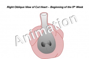
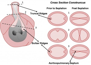
--Phoebe Norville 22:07, 28 September 2009 (EST) Content added:
--Mark Hill 13:56, 11 September 2009 (EST) Cardiac neural crest stem cells. Sieber-Blum M. Anat Rec A Discov Mol Cell Evol Biol. 2004 Jan;276(1):34-42. Review. PMID: 14699632
--Mark Hill 01:52, 10 September 2009 (EST) Truncus appears in the human embryo between Carnegie stages 12 to 13 The formation, septation and fate of the truncus arteriosus in man. F Orts-Llorca, J Puerta Fonolla, and J Sobrado J Anat. 1982 January; 134(Pt 1): 41–56. PMCID: PMC1167935
Background Reading
--Mark Hill 01:35, 10 September 2009 (EST)
- Septation and separation within the outflow tract of the developing heart Sandra Webb, Sonia R Qayyum, Robert H Anderson, Wouter H Lamers, and Michael K Richardson J Anat. 2003 April; 202(4): 327–342. doi: 10.1046/j.1469-7580.2003.00168.x. PMCID: PMC1571094
- Observations on the development of the aortico-pulmonary spiral septum in the mouse. K Fananapazir and M H Kaufman J Anat. 1988 June; 158: 157–172. PMCID: PMC1261986

