Scanning Electron Microscopy: Difference between revisions
From Embryology
| Line 3: | Line 3: | ||
==Introduction== | ==Introduction== | ||
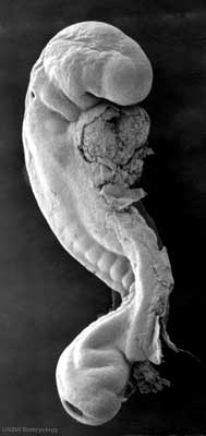
[[File:Stage12 sem1c.jpg|thumb|'''Human Embryo''' <br>(stage 12, week 4) SEM]] | [[File:Stage12 sem1c.jpg|thumb|'''Human Embryo''' <br>(stage 12, week 4) SEM]] | ||
The Scanning Electron Microscope (SEM) was a development of the electron microscope. Unlike a light microscope, using light, the electron microscope uses a focussed beam of electrons to image materials. | |||
On this site the acronym "SEM" is used to denote a '''S'''canning '''E'''lectron '''M'''icrograph, the image produced by this form of microscopy. | |||
:'''Links:''' [[:Category:Scanning EM|Category:Scanning EM]] | :'''Links:''' [[:Category:Scanning EM|Category:Scanning EM]] | ||
Revision as of 08:40, 1 June 2011
Notice - Mark Hill
Currently this page is only a template and will be updated (this notice removed when completed).Introduction
The Scanning Electron Microscope (SEM) was a development of the electron microscope. Unlike a light microscope, using light, the electron microscope uses a focussed beam of electrons to image materials.
On this site the acronym "SEM" is used to denote a Scanning Electron Micrograph, the image produced by this form of microscopy.
- Links: Category:Scanning EM
Glossary Links
- Glossary: A | B | C | D | E | F | G | H | I | J | K | L | M | N | O | P | Q | R | S | T | U | V | W | X | Y | Z | Numbers | Symbols | Term Link
Cite this page: Hill, M.A. (2024, May 2) Embryology Scanning Electron Microscopy. Retrieved from https://embryology.med.unsw.edu.au/embryology/index.php/Scanning_Electron_Microscopy
- © Dr Mark Hill 2024, UNSW Embryology ISBN: 978 0 7334 2609 4 - UNSW CRICOS Provider Code No. 00098G
