Quicktime Movie - Neural Sylvian Fissure: Difference between revisions
From Embryology
(Created page with '{| border='0px' |- | <Flowplayer height="355" width="355" autoplay="true">Neural_-_Sylvian_fissure.flv</Flowplayer> | valign="top" | This is an animation created from individual …') |
(Redirected page to Human Sylvian Fissure Movie) |
||
| (One intermediate revision by one other user not shown) | |||
| Line 1: | Line 1: | ||
#REDIRECT [[Human_Sylvian_Fissure_Movie]] | |||
{| border='0px' | {| border='0px' | ||
|- | |- | ||
| < | | <qt>file=Neural_-_Sylvian_fissure.mov|width=355px|height=385px|controller=true|autoplay=false</qt> | ||
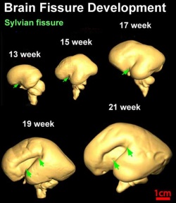
| valign="top" | This is an animation created from individual three-dimensional reconstruction of the lateral (top row) surface of 13–21 week brains to reveal the development of the Sylvian or lateral fissure (green arrow). | | valign="top" | This is an animation created from individual three-dimensional reconstruction of the lateral (top row) surface of 13–21 week brains to reveal the development of the Sylvian or lateral fissure (green arrow). | ||
| Line 7: | Line 8: | ||
|- | |- | ||
|} | |} | ||
==Reference== | ==Reference== | ||
| Line 15: | Line 15: | ||
Original File Name: Figure 5 Original image modified by scaling relative to 21 weeks, labeling, and reorganizing arrangement of image. | Original File Name: Figure 5 Original image modified by scaling relative to 21 weeks, labeling, and reorganizing arrangement of image. | ||
Latest revision as of 12:54, 8 March 2013
Redirect to:
| width=355px|height=385px|controller=true|autoplay=false</qt> | This is an animation created from individual three-dimensional reconstruction of the lateral (top row) surface of 13–21 week brains to reveal the development of the Sylvian or lateral fissure (green arrow). |
Reference
<pubmed>19339620</pubmed>| PMC2721010 | J Neurosci.
Original File Name: Figure 5 Original image modified by scaling relative to 21 weeks, labeling, and reorganizing arrangement of image.
