Magnetic Resonance Imaging: Difference between revisions
| Line 24: | Line 24: | ||
File:Cochlea_MRI_02.jpg | File:Cochlea_MRI_02.jpg | ||
</gallery> | </gallery> | ||
== References == | |||
<references/> | |||
===Reviews=== | |||
===Articles=== | |||
===Search PubMed=== | |||
'''Search Pubmed:''' [http://www.ncbi.nlm.nih.gov/sites/entrez?db=pubmed&cmd=search&term=Embryo%20Magnetic%20Resonance%20Imaging Embryo Magnetic Resonance Imaging] | [http://www.ncbi.nlm.nih.gov/sites/entrez?db=pubmed&cmd=search&term=Magnetic%20Resonance%20Imaging Magnetic Resonance Imaging] | | |||
{{Template:Glossary}} | |||
{{Template:Footer}} | |||
[[Category:Magnetic Resonance Imaging]] | [[Category:Magnetic Resonance Imaging]] | ||
Revision as of 11:05, 16 August 2010
Introduction
Recently there have been several groups preparing developmental embryo atlases of several species, including human, based upon imaging of different age embryos. There have also been studies of adult anatomical structures and the placenta.
Magnetic Resonance Imaging (MRI) began in 1977 and uses magnetism, radio waves, and a computer to produce images either as individual slices or reconstructed to give three dimensional (3D) views of specific anatomical regions or structures.
MRI can be used in fetuses at 18 weeks gestational age or later and has been used mainly in brain and spinal diagnosis, and has also been used to investigate other abnormalities of pregnancy. (More? BrighamRAD Twin Gestation with Complete Hydatidiform Mole)
About MRI
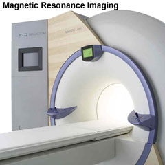
A strong magnetic field (up to 1.5 to 4 Tesla) is generated in the machine through which the body is passed (the centre of the "donut ring" seen in the above image). The earth's natural magnetic field is about 0.5 Gauss compared to 15,000 Gauss (1.5 Tesla) in the MRI.
Some Recent Findings
Adult Structures
Inner Ear
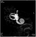
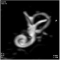
The 3D reconstructed technique was used to acquire coronal and axial images of the adult inner ear. The coronal section reconstruction was chosen since it increases visibility of the turns of the cochlea.[1]
References
- ↑ <pubmed>19575114</pubmed>| Braz J Otorhinolaryngol.
Reviews
Articles
Search PubMed
Search Pubmed: Embryo Magnetic Resonance Imaging | Magnetic Resonance Imaging |
Glossary Links
- Glossary: A | B | C | D | E | F | G | H | I | J | K | L | M | N | O | P | Q | R | S | T | U | V | W | X | Y | Z | Numbers | Symbols | Term Link
Cite this page: Hill, M.A. (2024, May 4) Embryology Magnetic Resonance Imaging. Retrieved from https://embryology.med.unsw.edu.au/embryology/index.php/Magnetic_Resonance_Imaging
- © Dr Mark Hill 2024, UNSW Embryology ISBN: 978 0 7334 2609 4 - UNSW CRICOS Provider Code No. 00098G


