File:Wen1928-Fig13.jpg
Original file (851 × 800 pixels, file size: 148 KB, MIME type: image/jpeg)
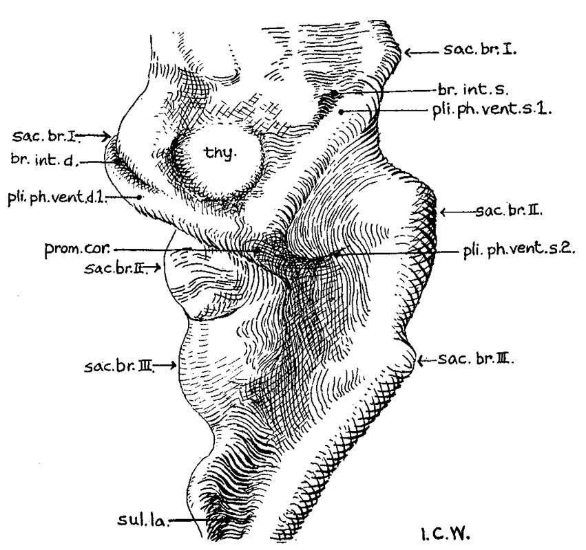
Fig. 13. A ventral View of a wax model of the pharynx of the 17 Somite embryo H951
X 100.
The external configuration of the ventral pharyngeal wall is shown in figure 13. In many respects it resembles the pharynx of Grosser’s 15—somite embryo, Halg ( ’11, his fig. 10, pl. 2), which is in turn similar to that of Davis’ 20-somite specimen. The thyroid anlage represented by a prominent swelling is situated a little to the right side rather than in the median plane. Caudal to it, but to the midline, there is another prominent swelling, which Davis has called the ‘heart swelling’, as produced by “the molding of the pharyngeal wall in the notch between the ventral aorta and the heart atrium” (’23, p. 25). In the 17—somite embryo we find that this swelling involves a dorsoventral expansion of the pharyngeal cavity at the level just caudal to the ventral aortic trunk. From it a pair of folds (ventral pharyngeal folds) proceeds obliquely cephalolaterally to the ventral angle of each first visceral pouch, marking off this clearly from the second pouch. Similarly, a second pair of folds, but much shorter than the first, can be traced from the heart swelling laterally toward the ventral angle of the second visceral pouch, more clearly marked on the left than on the right side of the embryo. Caudal to the second pair of folds, the heart swelling gradually fades out, and no more such folds can be found. At the cephalic end of each. first Ventral pharyngeal fold there is an invagination of the pharyngeal floor into the visceral pouch. This is the ‘Kiemenrudiment’ (br.mt.md.) of Grosser (’11, compare his figs. 4, 7, and 10), to which we shall come presently. Caudal to the level of the second Visceral pouch, the pharynx becomes narrowed down and compressed dorsoventrally with a concavity directed dorsally. Laterally, the first two pouches are in contact with the ectoderm, but the wall caudal to the second visceral pouch only indicates the impending extension of the third, which at this stage has not as yet reached its ectodermal area
| Abbreviations for all Figures | |
|---|---|
|
|
| Historic Disclaimer - information about historic embryology pages |
|---|
 |
- Links: fig 1 | Plate 2 | fig 2 | fig 3 | fig 4 | fig 5 | fig 6 | fig 7 | fig 8 | fig 9 | fig 10 | fig 11 | fig 12 | fig 13 | fig 14 | fig 15 | fig 16 | fig 17 | fig 18 | fig 19 | fig 21 | fig 21 | fig 22 | fig 23 | fig 24 | fig 25 | fig 26 | fig 27 | fig 28 | fig 29 | Wen 1928 | Carnegie stage 11 | Carnegie stage 12 | Historic Papers
Reference
Wen IC. The anatomy of human embryos with seventeen to twenty-three pairs of somites (1928) J. Comp. Neural., 45: 301-376.
Cite this page: Hill, M.A. (2024, April 27) Embryology Wen1928-Fig13.jpg. Retrieved from https://embryology.med.unsw.edu.au/embryology/index.php/File:Wen1928-Fig13.jpg
- © Dr Mark Hill 2024, UNSW Embryology ISBN: 978 0 7334 2609 4 - UNSW CRICOS Provider Code No. 00098G
File history
Click on a date/time to view the file as it appeared at that time.
| Date/Time | Thumbnail | Dimensions | User | Comment | |
|---|---|---|---|---|---|
| current | 15:46, 20 April 2016 | | 851 × 800 (148 KB) | Z8600021 (talk | contribs) | |
| 15:46, 20 April 2016 |  | 1,312 × 1,094 (257 KB) | Z8600021 (talk | contribs) |
You cannot overwrite this file.
File usage
The following page uses this file: