File:Wen1928-Fig07.jpg

Original file (1,265 × 648 pixels, file size: 166 KB, MIME type: image/jpeg)
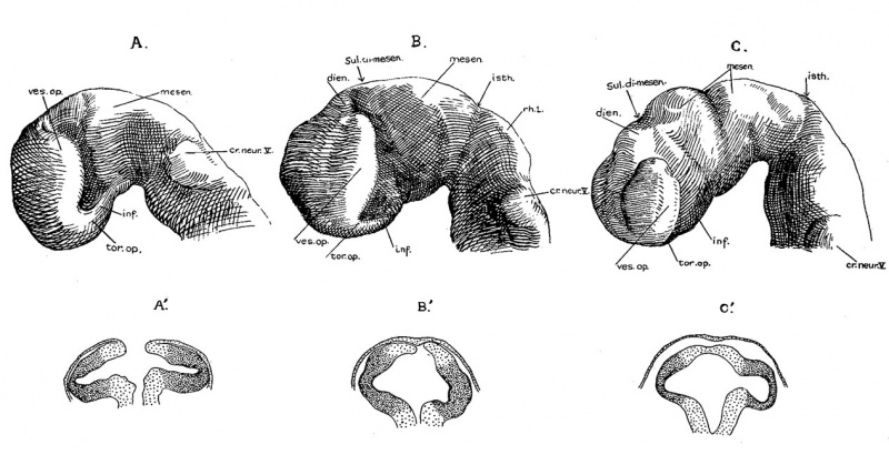
Fig. 7 Lateral Views of the Brain
A, 16—somite C.C.470;
B, 17~somite H951;
C, 22—somite H1093—all drawn from models and reduced to a magnification of 66% diameters. Note the absolute decrease in the size of the optic vesicle and the relations of the midbrain to the cranial fiexnre.
A’, B’. C’, are sections (nos. 28, 25, and 34, respectively) taken from the corresponding embryos and magnified 66% times. The region which in external view appears as optic vesicle is deeply stippled.
Externally, the optic vesicle first appears to spring from the greater part of the forebrain (fig. 7, A) and extends laterally, dorsally, and slightly caudad. Soon each vesicle is marked off from the forebrain wall by a deeper, caudally located groove and a shallow rostral sulcus (fig. 7, B). With the separation of the vesicle from the brain wall, these grooves become ventral and dorsal in position with reference to the forebrain, but not the embryo as a whole, as may be seen in figure 7, 0. They may accordingly be termed ventral and dorsal limiting sulci, respectively. In the 17 somite embryo the more caudal ventral sulcus, beginning almost at the torus, extends dorsally behind the optic vesicle and has begun to arch over it to become continuous with the dorsal sulcus which fades out at about the middle of the vesicle. From the ventricular surface we see (fig. 4) the optic ventricle opening into the third ventricle by a large oval orifice, bounded caudally (ventrally) by a strongly indented ridge which corresponds to the ventral limiting sulcus.
| Abbreviations for all Figures | |
|---|---|
|
|
| Historic Disclaimer - information about historic embryology pages |
|---|
 |
- Links: fig 1 | Plate 2 | fig 2 | fig 3 | fig 4 | fig 5 | fig 6 | fig 7 | fig 8 | fig 9 | fig 10 | fig 11 | fig 12 | fig 13 | fig 14 | fig 15 | fig 16 | fig 17 | fig 18 | fig 19 | fig 21 | fig 21 | fig 22 | fig 23 | fig 24 | fig 25 | fig 26 | fig 27 | fig 28 | fig 29 | Wen 1928 | Carnegie stage 11 | Carnegie stage 12 | Historic Papers
Reference
Wen IC. The anatomy of human embryos with seventeen to twenty-three pairs of somites (1928) J. Comp. Neural., 45: 301-376.
Cite this page: Hill, M.A. (2024, April 27) Embryology Wen1928-Fig07.jpg. Retrieved from https://embryology.med.unsw.edu.au/embryology/index.php/File:Wen1928-Fig07.jpg
- © Dr Mark Hill 2024, UNSW Embryology ISBN: 978 0 7334 2609 4 - UNSW CRICOS Provider Code No. 00098G
File history
Click on a date/time to view the file as it appeared at that time.
| Date/Time | Thumbnail | Dimensions | User | Comment | |
|---|---|---|---|---|---|
| current | 14:07, 20 April 2016 | | 1,265 × 648 (166 KB) | Z8600021 (talk | contribs) | |
| 14:03, 20 April 2016 |  | 1,280 × 781 (215 KB) | Z8600021 (talk | contribs) | Wen, I. C., The anatomy of human embryos with seventeen to twenty-three pairs of somites J. Comp. Neural, 1928, 45:301-376. Historic Embryology Papers {| clas... |
You cannot overwrite this file.
File usage
The following page uses this file: