File:Waterston19.jpg
From Embryology
Size of this preview: 467 × 600 pixels. Other resolution: 500 × 642 pixels.
Original file (500 × 642 pixels, file size: 66 KB, MIME type: image/jpeg)
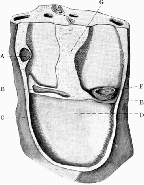
Fig. 19. Model after removal of the heart.
Wax-plate reconstruction model in fig.17, after removal of the heart.
Legend
- A, Right duct. of Cuvier
- B, sinus venosus
- C, body wall
- D, floor of pericardium (septum trans- versum)
- E, left duct of Cuvier
- F, left portion of atrium
- G, dorsal mesocardium. The pericardio-peritorieal connexions lie behind B and medial to F respectively.
Carnegie Staging Comparison: A 27 somite stage embryo would be similar to a Carnegie stage 12 (26 - 30 days), caudal neuropore closes, Somite Number 21-29.
| Historic Disclaimer - information about historic embryology pages |
|---|
 |
- Historic Paper Links: 13-14 Somites | 22 Somites | 23 Somites | 25 Somites | 27 Somites | Mall Human Embryo Collection | Embryology History | Carnegie stage 11 | Carnegie stage 12 | Journal of Anatomy | Embryonic Development | Category:Historic Embryology
Reference
<pubmed>17233016</pubmed>| PMC1288995
File history
Click on a date/time to view the file as it appeared at that time.
| Date/Time | Thumbnail | Dimensions | User | Comment | |
|---|---|---|---|---|---|
| current | 14:03, 27 January 2012 | | 500 × 642 (66 KB) | S8600021 (talk | contribs) | ==Fig. 19. Model after removal of the heart.== Wax-plate reconstruction model in fig.17, after removal of the heart. ===Legend=== * A, Right duct. of Cuvier * B, sinus venosus * C, body wall * D, floor of pericardium (septum trans- versum) * E, left du |
You cannot overwrite this file.