File:Ventricular septal defect 01.jpg: Difference between revisions
From Embryology
mNo edit summary |
|||
| Line 1: | Line 1: | ||
==Ventricular Septal Defect (ultrasound)== | ==Ventricular Septal Defect (ultrasound)== | ||
There is a defect in the ventricular septum adjacent to the atrioventricular valves. Blood flow is seen across the defect on | There is a defect in the ventricular septum adjacent to the atrioventricular valves. Blood flow is seen across the defect on Colour Doppler imaging. | ||
{{Stanley Ng}} | |||
{{Footer}} | {{Footer}} | ||
[[Category:Cardiovascular]] [[Category:Heart]] [[Category:Abnormal Development]] | [[Category:Cardiovascular]] [[Category:Heart]] [[Category:Abnormal Development]] | ||
Revision as of 17:50, 22 June 2016
Ventricular Septal Defect (ultrasound)
There is a defect in the ventricular septum adjacent to the atrioventricular valves. Blood flow is seen across the defect on Colour Doppler imaging.
Dr Stanley Ng - Obstetrical and gynecological sonologist (Sydney) for providing fetal ultrasound images and movie clips.
Cite this page: Hill, M.A. (2024, April 30) Embryology Ventricular septal defect 01.jpg. Retrieved from https://embryology.med.unsw.edu.au/embryology/index.php/File:Ventricular_septal_defect_01.jpg
- © Dr Mark Hill 2024, UNSW Embryology ISBN: 978 0 7334 2609 4 - UNSW CRICOS Provider Code No. 00098G
File history
Click on a date/time to view the file as it appeared at that time.
| Date/Time | Thumbnail | Dimensions | User | Comment | |
|---|---|---|---|---|---|
| current | 17:46, 22 June 2016 | 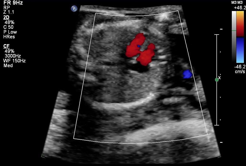 | 1,024 × 692 (84 KB) | Z8600021 (talk | contribs) |
You cannot overwrite this file.
File usage
The following 2 pages use this file: