File:Uterine gland secretory phase.jpg: Difference between revisions
From Embryology
No edit summary |
No edit summary |
||
| Line 10: | Line 10: | ||
Image Source: UWA Blue Histology | Image Source: UWA Blue Histology | ||
http://www.lab.anhb.uwa.edu.au/mb140/CorePages/FemaleRepro/femalerepro.htm#Uterus | http://www.lab.anhb.uwa.edu.au/mb140/CorePages/FemaleRepro/femalerepro.htm#Uterus | ||
[[Category:Histology]] [[Category:Genital]] [[Category:Uterus]] | |||
Revision as of 21:39, 13 October 2009
Uterine gland secretory phase
Histology showing endometrium uterine glands and surrounding stromal cells during the secretory phase of menstrual cycle. Note the expanded gland lumen and relative amount of glandular and stromal cells.
Compare this image with the menstrual proliferative phase images.
H&E stain
Image Source: UWA Blue Histology http://www.lab.anhb.uwa.edu.au/mb140/CorePages/FemaleRepro/femalerepro.htm#Uterus
File history
Click on a date/time to view the file as it appeared at that time.
| Date/Time | Thumbnail | Dimensions | User | Comment | |
|---|---|---|---|---|---|
| current | 12:55, 2 February 2012 |  | 400 × 533 (49 KB) | S8600021 (talk | contribs) | increase image size |
| 10:24, 3 August 2009 | 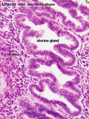 | 300 × 400 (65 KB) | MarkHill (talk | contribs) | Uterine gland secretory phase |
You cannot overwrite this file.
File usage
The following 11 pages use this file: