File:Trilaminar embryo.jpg: Difference between revisions
No edit summary |
mNo edit summary |
||
| (4 intermediate revisions by the same user not shown) | |||
| Line 2: | Line 2: | ||
The embryonic disc has been broken to expose the early three germ layers that form the entire embryo (scanning electron micrograph). | |||
# Ectoderm | # [[Ectoderm]] - columnar epithelium | ||
# Mesoderm | # [[Mesoderm]] - embryonic connective tissue (mesenchyme) | ||
# Endoderm | # [[Endoderm]] - cuboidal epithelium | ||
{{ | * The amniotic cavity would lie above the ectoderm layer. | ||
* The yolk sac would initially lie below the endoderm layer, later this would be the gastrointestinal tract within the embryo. | |||
:Links: [[Gastrulation]] | [[Ectoderm]] | [[Mesoderm]] | [[Endoderm]] | [[Week 3]] | |||
---- | |||
{{SEM}} | |||
{{Footer}} | |||
[[Category:Electron Micrograph]] [[Category:Week 3]] | [[Category:Electron Micrograph]] [[Category:Week 3]] | ||
Latest revision as of 09:45, 5 May 2014
Trilaminar Embryo (SEM)
The embryonic disc has been broken to expose the early three germ layers that form the entire embryo (scanning electron micrograph).
- Ectoderm - columnar epithelium
- Mesoderm - embryonic connective tissue (mesenchyme)
- Endoderm - cuboidal epithelium
- The amniotic cavity would lie above the ectoderm layer.
- The yolk sac would initially lie below the endoderm layer, later this would be the gastrointestinal tract within the embryo.
- Links: Gastrulation | Ectoderm | Mesoderm | Endoderm | Week 3
Image Source: Scanning electron micrographs of the Carnegie stages of the early human embryos are reproduced with the permission of Prof Kathy Sulik, from embryos collected by Dr. Vekemans and Tania Attié-Bitach. Images are for educational purposes only and cannot be reproduced electronically or in writing without permission.
Cite this page: Hill, M.A. (2024, May 5) Embryology Trilaminar embryo.jpg. Retrieved from https://embryology.med.unsw.edu.au/embryology/index.php/File:Trilaminar_embryo.jpg
- © Dr Mark Hill 2024, UNSW Embryology ISBN: 978 0 7334 2609 4 - UNSW CRICOS Provider Code No. 00098G
File history
Click on a date/time to view the file as it appeared at that time.
| Date/Time | Thumbnail | Dimensions | User | Comment | |
|---|---|---|---|---|---|
| current | 13:41, 23 April 2010 | 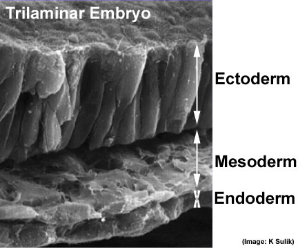 | 432 × 359 (32 KB) | S8600021 (talk | contribs) | Trilaminar embryo (SEM) {{Template:SEM}} |
You cannot overwrite this file.
File usage
The following 29 pages use this file:
- 2010 BGD Lecture - Development of the Embryo/Fetus 1
- 2010 BGD Practical 3 - Gastrulation
- 2010 Foundations Lecture - Introduction to Human Development
- 2010 Lab 2
- 2010 Lecture 5
- 2011 Lab 2 - Week 3
- 2011 Lab 6 - Trilaminar Embryo
- ANAT2341 Lab 6 - Trilaminar Embryo
- BGDA Lecture - Development of the Embryo/Fetus 1
- BGDA Lecture - Development of the Embryo/Fetus 2
- BGDA Practical 3 - Gastrulation
- BGDA Practical 7 - Week 3
- BGDB Face and Ear - Trilaminar Embryo
- BGDB Gastrointestinal - Activity 1
- BGDB Gastrointestinal - Trilaminar Embryo
- BGD Lecture - Gastrointestinal System Development
- E
- Ectoderm
- Endoderm
- Foundations Lecture - Introduction to Human Development
- Foundations Practical - Week 3 and 4
- Human Embryo SEM
- Lecture - Ectoderm Development
- Lecture - Mesoderm Development
- Lecture - Week 3 Development
- Mesoderm
- Pre-Medicine Program - Embryology
- REI - Reproductive Medicine Seminar 2018
- Royal Hospital for Women - Reproductive Medicine Seminar 2018