File:Tooth development stage.jpg: Difference between revisions
No edit summary |
|||
| (7 intermediate revisions by 2 users not shown) | |||
| Line 7: | Line 7: | ||
'''(C)''' At the sites of the dental placodes the epithelial cells proliferate and intrude within the mesenchyme forming the tooth buds. At this developmental stage the odontogenic potential is lost form the epithelium and granted to the ectomesenchyme. | '''(C)''' At the sites of the dental placodes the epithelial cells proliferate and intrude within the mesenchyme forming the tooth buds. At this developmental stage the odontogenic potential is lost form the epithelium and granted to the ectomesenchyme. | ||
'''(D)''' The bud folds in and acquires initially the form of an inverted cap and later the form of a bell | '''(D)''' The bud folds in and acquires initially the form of an inverted cap and later the form of a bell. | ||
'''(E)''' cl = cervical loop, iee = inner enamel epithelium, oee = outer enamel epithelium, pek = primary enamel knot, sek = secondary enamel knots. | '''(E)''' cl = cervical loop, iee = inner enamel epithelium, oee = outer enamel epithelium, pek = primary enamel knot, sek = secondary enamel knots. | ||
:"The ancestor of recent vertebrate teeth was a tooth-like structure on the outer body surface of jawless fishes. Over the course of 500,000,000 years of evolution, many of those structures migrated into the mouth cavity. In addition, the total number of teeth per dentition generally decreased and teeth morphological complexity increased. Teeth form mainly on the jaws within the mouth cavity through mutual, delicate interactions between dental epithelium and oral ectomesenchyme. These interactions involve spatially restricted expression of several, teeth-related genes and the secretion of various transcription and signaling factors." | |||
: | {{Tooth stages}} | ||
===Reference=== | |||
{{#pmid:19266065}} | |||
====Copyright==== | |||
© Ivyspring International Publisher. This is an open-access article distributed under the terms of the Creative Commons License (http://creativecommons.org/licenses/by-nc-nd/3.0/). Reproduction is permitted for personal, noncommercial use, provided that the article is in whole, unmodified, and properly cited. | |||
Adapted from original file name: Ijbsv05p0226g04.jpg http://www.pubmedcentral.nih.gov/articlerender.fcgi?artid=2651620&rendertype=figure&id=F4 | |||
[[Category:Tooth]] | {{Footer}} | ||
[[Category:Tooth]] [[Category:Integumentary]] | |||
Latest revision as of 14:32, 4 February 2019
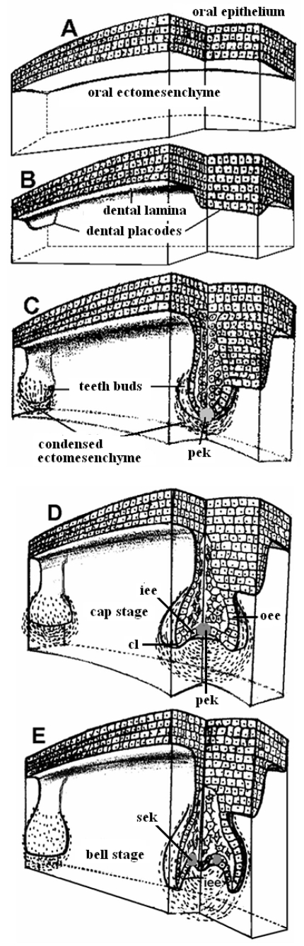
Stages in Tooth Development
(A) Pre-patterned oral ectoderm is in close contact with cranial, neural crest ectomesenchyme. At this stage (ED 10) the odontogenic potential resides in the epithelium.
(B) The epithelial cells secrete specific signals in different areas, proliferate and form a band of epithelial tissue, the dental lamina and the dental placodes.
(C) At the sites of the dental placodes the epithelial cells proliferate and intrude within the mesenchyme forming the tooth buds. At this developmental stage the odontogenic potential is lost form the epithelium and granted to the ectomesenchyme.
(D) The bud folds in and acquires initially the form of an inverted cap and later the form of a bell.
(E) cl = cervical loop, iee = inner enamel epithelium, oee = outer enamel epithelium, pek = primary enamel knot, sek = secondary enamel knots.
- "The ancestor of recent vertebrate teeth was a tooth-like structure on the outer body surface of jawless fishes. Over the course of 500,000,000 years of evolution, many of those structures migrated into the mouth cavity. In addition, the total number of teeth per dentition generally decreased and teeth morphological complexity increased. Teeth form mainly on the jaws within the mouth cavity through mutual, delicate interactions between dental epithelium and oral ectomesenchyme. These interactions involve spatially restricted expression of several, teeth-related genes and the secretion of various transcription and signaling factors."
- Tooth stages: lamina | placode stage | bud stage | cap stage | bell stage | all stages | Tooth Development
Reference
Koussoulakou DS, Margaritis LH & Koussoulakos SL. (2009). A curriculum vitae of teeth: evolution, generation, regeneration. Int. J. Biol. Sci. , 5, 226-43. PMID: 19266065
Copyright
© Ivyspring International Publisher. This is an open-access article distributed under the terms of the Creative Commons License (http://creativecommons.org/licenses/by-nc-nd/3.0/). Reproduction is permitted for personal, noncommercial use, provided that the article is in whole, unmodified, and properly cited.
Adapted from original file name: Ijbsv05p0226g04.jpg http://www.pubmedcentral.nih.gov/articlerender.fcgi?artid=2651620&rendertype=figure&id=F4
Cite this page: Hill, M.A. (2024, April 27) Embryology Tooth development stage.jpg. Retrieved from https://embryology.med.unsw.edu.au/embryology/index.php/File:Tooth_development_stage.jpg
- © Dr Mark Hill 2024, UNSW Embryology ISBN: 978 0 7334 2609 4 - UNSW CRICOS Provider Code No. 00098G
File history
Click on a date/time to view the file as it appeared at that time.
| Date/Time | Thumbnail | Dimensions | User | Comment | |
|---|---|---|---|---|---|
| current | 09:25, 9 August 2009 | 430 × 1,338 (303 KB) | S8600021 (talk | contribs) | Stages in teeth development (A) Pre-patterned oral ectoderm is in close contact with cranial, neural crest ectomesenchyme. At this stage (ED 10) the odontogenic potential resides in the epithelium. (B) The epithelial cells secrete specific signals in d |
You cannot overwrite this file.
File usage
The following page uses this file: