File:Thomson1919 fig03.jpg

Original file (1,226 × 1,110 pixels, file size: 308 KB, MIME type: image/jpeg)
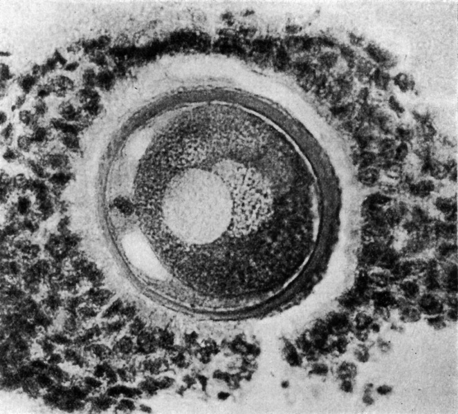
Fig. 3. Human ovum in discus proligerus in anaphase of first division and commencement of metaphase of second division
x 600.
Ovum in discus proligerus in Graafian follicle. Thickness of section 0.01 mm, stained with Weigert’s Iron Haematoxylin and Van Giesen. Size including Zona 0.097 x 0.088 mm; inside Zona 0.083 x 0.08 mm.
From a woman whose ovary was forwarded without any history, in the same case a morula and an ovarian blastula were found; these will be described in a subsequent paper. In this case the Fallopian tubes were much twisted and bound down by adhesions——presumably impervious to the descent of an ovum though apparently permitting the passage of spermatozoa.
The section here displays a more spherical appearance.
The cells of the Corona radiata are neither so discrete nor so clearly seen as in the previous figures (1 and 2). They are separated from the outer surface of the Zona pellucida by a zone about 0.001 mm in width, which exhibits but faint indications of structure, the fibrillar arcades being conspicuous by their absence and the tissue intervening, between the outer surface of the Zona pellucida and the innermost row of the cells of the Corona radiata on one side of the egg, appears but faintly striated and granular, whereas on the opposite side of the ovum the interval appears clearer as if occupied with fluid—further, there is a breach in the continuity of the girdle of surrounding coronal cells, as if by the penetration of the liquor folliculi, a stage, possibly, in the liberation of the oocyte from the cells of the discus proligerus immediately around it. It may reasonably be assumed that the arcading of the tissue connecting the cells of the Corona radiata with the Zona pellucida exhibited in the previous figures is part of the same process, brought about by the permeation of fluid (liquor folliculi?) in between.
The Zona pellucida on the two sides of the egg exhibits a marked difference in section, varying from 0.007 mm on one side to 0.003 mm on the opposite side of the ovum, a difference due no doubt to the obliquity of the section.
The innermost, more homogeneous and ovular (‘?) layer of the Zona pellucida alone remains, the external fibrillar or ovarian layer having now all but disappeared on one side, whereas on the other side it is reduced to a faint reticulum. The Zona pellucida left exhibits traces of concentric lamination with a suggestion at one point of cellular structure. In this specimen I can see no evidence of radial striation.
File history
Click on a date/time to view the file as it appeared at that time.
| Date/Time | Thumbnail | Dimensions | User | Comment | |
|---|---|---|---|---|---|
| current | 13:49, 6 August 2015 |  | 1,226 × 1,110 (308 KB) | Z8600021 (talk | contribs) | |
| 13:48, 6 August 2015 |  | 1,443 × 1,306 (468 KB) | Z8600021 (talk | contribs) | ==Fig. 3. Human ovum in discus proligerus in anaphase of first division and commencement of metaphase of second division== x 600. Ovum in discus proligerus in Graafian follicle. Thickness of section 0.01 mm, stained with Weigert’s Iron Haematoxylin... |
You cannot overwrite this file.
File usage
The following page uses this file: