File:Testis histology.jpg
From Embryology
No higher resolution available.
Testis_histology.jpg (400 × 500 pixels, file size: 54 KB, MIME type: image/jpeg)
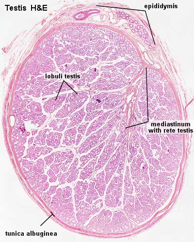
Testis, young and mature
(Stain - Haematoxylin Eosin)
- Use the lowest magnification available.
- Identify the capsule and the connective tissue septa extending from it.
- Identify lobules, convoluted seminiferous tubules and clusters of interstitial cells.
- The mediastinum testis and rete testis are not visible in all sections.
Links: Histology | Histology Stains | Blue Histology images copyright Lutz Slomianka 1998-2009. The literary and artistic works on the original Blue Histology website may be reproduced, adapted, published and distributed for non-commercial purposes. See also the page Histology Stains.
Cite this page: Hill, M.A. (2024, April 26) Embryology Testis histology.jpg. Retrieved from https://embryology.med.unsw.edu.au/embryology/index.php/File:Testis_histology.jpg
- © Dr Mark Hill 2024, UNSW Embryology ISBN: 978 0 7334 2609 4 - UNSW CRICOS Provider Code No. 00098G
File history
Click on a date/time to view the file as it appeared at that time.
| Date/Time | Thumbnail | Dimensions | User | Comment | |
|---|---|---|---|---|---|
| current | 16:26, 21 September 2009 | | 400 × 500 (54 KB) | S8600021 (talk | contribs) | |
| 16:25, 21 September 2009 | 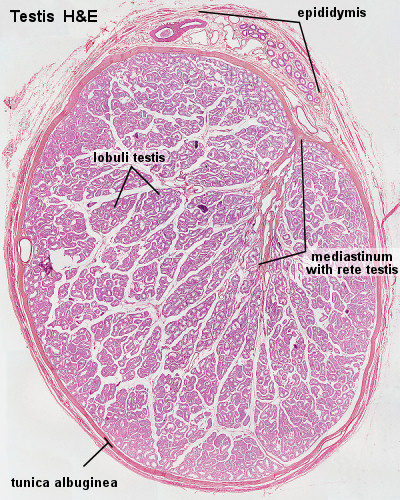 | 400 × 500 (158 KB) | S8600021 (talk | contribs) | Testis, young and mature - H&E Use the lowest magnification available. Identify the capsule and the connective tissue septa extending from it. Identify lobules, convoluted seminiferous tubules and clusters of interstitial cells. The mediastinum testis an |
You cannot overwrite this file.