File:Stricht-plate01.jpg
From Embryology

Size of this preview: 462 × 599 pixels. Other resolution: 1,156 × 1,500 pixels.
Original file (1,156 × 1,500 pixels, file size: 243 KB, MIME type: image/jpeg)
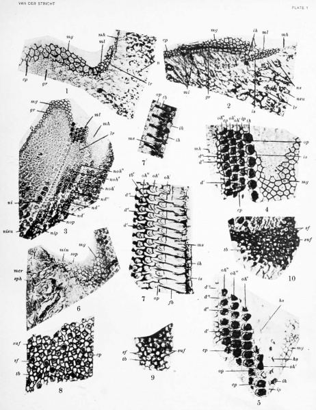
Plate 1
- Photograph of transverse section throughi tympanic wall of cochlea near apex. New-born dog. Fixation, trichloracetic acid; stain, iron hematoxylin, Stain - Congo red.
- Photograph of transverse section through second turn of spiral duct. Pig embryo 93. r> mm. Trichloracetic acid; iron hematoxylin, Stain - Congo red.
- Photograph of transverse, slightly oblique section through the greater and lesser ridges of second turn of cochlea, New-born dog. Trichloracetic acid; iron hematoxylin, Stain - Congo red.
- Photograph of section tangential to surface of the two epithelial ridges in second turn of cochlea. New-born dog. Trichloracetic acid; iron hematoxylin, Stain - Congo red.
- Photograph of section tangential to surface of the two epithelial riilges between second and third turn of cochlea. New-born dog. Trichloracetic acid; iron hematoxylin, Stain - Congo red.
- Photograph of section tangential to surface of crista spiralis and the greater epithelial ridge. Pig embryo 137 mm. Bouin's fluid; iron heraatoxylin, Stain - Congo red.
- 7'. Photographs of a section tangential to the surface of the organ of Corti in the adult bat (Vesperlilio fuscus) Bouin's fluid; iron hematoxylin, Stain - Congo red.
- Photograph of section tangential to surface of crista acustica. New-born dog. Trichloracetic acid; iron hematoxylin, Stain - Congo red.
- Photograph of section tangential to surface of crista acustica in adult bat (Vesperlilio fuscus). Zenker's fluid ; iron hematoxylin, Stain - Congo red.
- Photograph of section tangential to the surface of crista acustica in adult white rat. Trichloracetic acid; iron hematoxylin, Stain - Congo red.
- Stricht Links: Plate 1 | Plate 2 | Plate 3 | Plate 4 | Membrana Tectoria and the Crista Spiralis | Contributions to Embryology Series
| Historic Disclaimer - information about historic embryology pages |
|---|
 |
Reference
O. Van der Stricht. The Genesis and Structure of the Membrana Tectoria and the Crista Spiralis of the Cochlea. Contributions to Embryology (1918) Vol. 7 pp55- 86.
Cite this page: Hill, M.A. (2024, April 27) Embryology Stricht-plate01.jpg. Retrieved from https://embryology.med.unsw.edu.au/embryology/index.php/File:Stricht-plate01.jpg
- © Dr Mark Hill 2024, UNSW Embryology ISBN: 978 0 7334 2609 4 - UNSW CRICOS Provider Code No. 00098G
File history
Click on a date/time to view the file as it appeared at that time.
| Date/Time | Thumbnail | Dimensions | User | Comment | |
|---|---|---|---|---|---|
| current | 18:46, 7 April 2012 | | 1,156 × 1,500 (243 KB) | Z8600021 (talk | contribs) | |
| 08:45, 7 April 2011 | 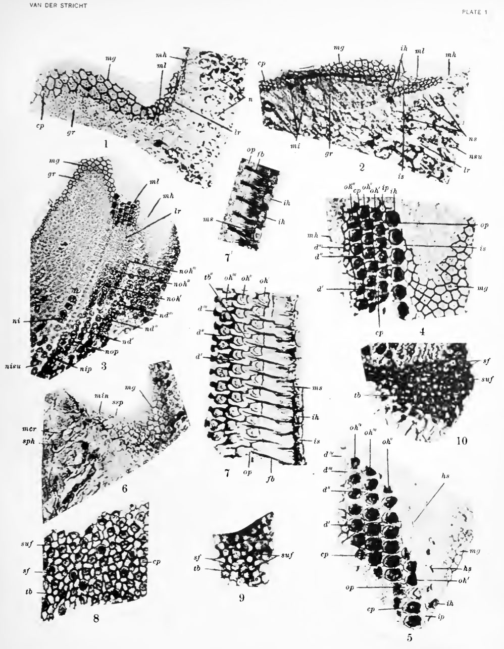 | 998 × 1,286 (284 KB) | S8600021 (talk | contribs) | ==Plate 1== 1. Photograph of transverse section throughi tympanic wall of cochlea near apex. New-born dog. Fixation, trichloracetic acid; stain, iron hematoxylin, Congo red. 2. Photograph of transverse section through second turn of spiral duct. Pig em |
You cannot overwrite this file.
File usage
The following page uses this file: