File:Streeter1919-fig02.jpg

Original file (2,023 × 1,318 pixels, file size: 499 KB, MIME type: image/jpeg)
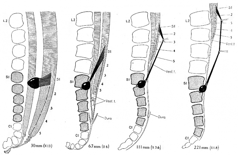
Fig. 2. Topographical relations of the caudal end of the spinal cord in the human fetus from the eighth to the twenty-fifth week
Comparison of these stages shows the rate and extent of caudal displacement of the vertebral column, in relation to the telzninal ventricle and the attachment of the sacral nerve roots, which constitute definite and fixed points on the spinal cord. In each case the dorsal root of the first sacral nerve is drawn in, and the attachment of the dorsal roots of the other sacral nerves is shown by heavy lines. The twenty-fifth to twenty-ninth vertebrae were regarded as sacral vertebrae, irrespective of form, as shown in stipple. The fetuses are listed in the Carnegie Collection follows: No. 75, 30 mm.; No. 1656, 67 mm.; No. Template:CE1673, 111 mm; N0. Template:CE1345, 221 mm. The first two are from profile reconstructions, the last two from dissections.
In figure 2 the specimens are enlarged upon a decreasing scale of magnification according to age, so that the segments of the different stages are brought to about the same size. This has been done in order to facilitate the comparison of segment levels. The actual elongation of the spinal root of a given nerve is greater, therefore, than would appear from the figure. Measurements of the dorsal root of the first sacral nerve from the margin of the ganglion to the point of entrance into the cord yield the following figures: 30-mm. fetus, 0.65 mm. long; 67-mm. fetus, 4.75 mm. long; 111-mm. fetus, 12.25 mm. long; 221-mm. fetus, 32 mm. long.
| Historic Disclaimer - information about historic embryology pages |
|---|
 |
Reference
Streeter GL. Factors involved in the formation of the filum terminale. (1919) Amer. J Anat. 22(1): 1-11.
Cite this page: Hill, M.A. (2024, April 27) Embryology Streeter1919-fig02.jpg. Retrieved from https://embryology.med.unsw.edu.au/embryology/index.php/File:Streeter1919-fig02.jpg
- © Dr Mark Hill 2024, UNSW Embryology ISBN: 978 0 7334 2609 4 - UNSW CRICOS Provider Code No. 00098G
File history
Click on a date/time to view the file as it appeared at that time.
| Date/Time | Thumbnail | Dimensions | User | Comment | |
|---|---|---|---|---|---|
| current | 23:53, 11 September 2015 | | 2,023 × 1,318 (499 KB) | Z8600021 (talk | contribs) | |
| 23:44, 11 September 2015 | 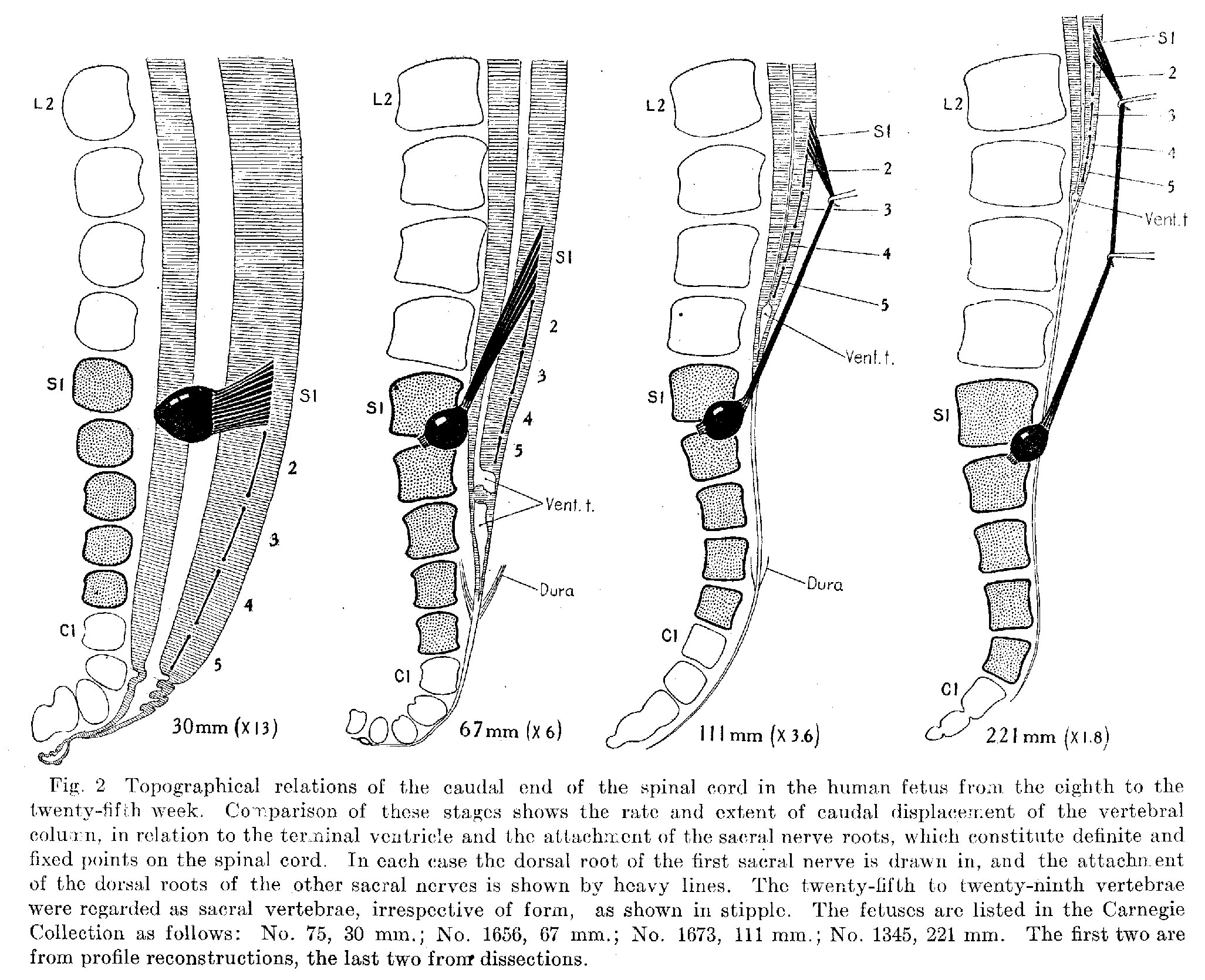 | 2,053 × 1,670 (693 KB) | Z8600021 (talk | contribs) |
You cannot overwrite this file.
File usage
The following 2 pages use this file: