File:Stage 22 image 214.jpg
From Embryology
Stage_22_image_214.jpg (738 × 554 pixels, file size: 203 KB, MIME type: image/jpeg)
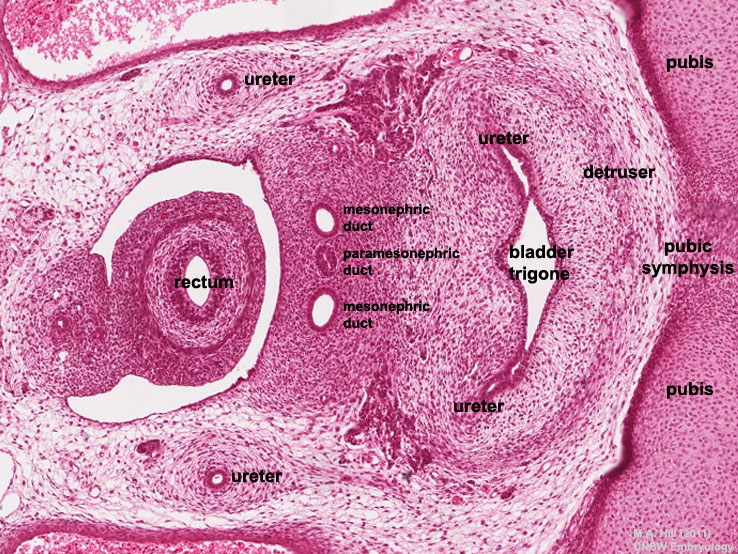
Developing Pelvic Region - Human Embryo Carnegie stage 22
- bladder (trigone)
- ureters
- mesonephric duct
- paramesonephric duct
- gastrointestinal tract (rectum)
- mesentery
- placental blood vessels (placental arteries)
- pelvis (pubic symphysis)
- axial skeleton
- Links: Carnegie stage 22 | Carnegie stage 22 | unlabeled | labeled | large 1200px | G7 urogenital
- Related Images: large 1200px | large 1000px | medium 800px | small 400px
| Selected Embryo Histology - Week 8 (Stage 22) |
|---|
|
| Links: Carnegie stage 22 | Week 8 |
Image Source: UNSW Embryology, no reproduction without permission.
Cite this page: Hill, M.A. (2024, April 27) Embryology Stage 22 image 214.jpg. Retrieved from https://embryology.med.unsw.edu.au/embryology/index.php/File:Stage_22_image_214.jpg
- © Dr Mark Hill 2024, UNSW Embryology ISBN: 978 0 7334 2609 4 - UNSW CRICOS Provider Code No. 00098G
File history
Click on a date/time to view the file as it appeared at that time.
| Date/Time | Thumbnail | Dimensions | User | Comment | |
|---|---|---|---|---|---|
| current | 22:23, 5 May 2011 |  | 738 × 554 (203 KB) | S8600021 (talk | contribs) | ==Developing Pelvic Region - Human Embryo Carnegie stage 22== * bladder (trigone) * ureters * mesonephric duct * paramesonephric duct * gastrointestinal tract (rectum) * mesentery * placental blood vessels * pelvis (pubic symphysis) * axial skeleton :' |
You cannot overwrite this file.
File usage
The following 45 pages use this file:
- B
- BGDA Practical 7 - Week 8
- BGDB Sexual Differentiation - Late Embryo
- BGD Lecture - Sexual Differentiation
- Carnegie stage 22
- Developmental Signals - Anti-Mullerian Hormone
- Gastrointestinal Tract - Intestine Development
- Gastrointestinal Tract - Mesentery Development
- Historic Embryology Vignette
- REI - Reproductive Medicine Seminar 2018
- Royal Hospital for Women - Reproductive Medicine Seminar 2018
- Urinary Bladder Development
- File:Johannes Muller.jpg
- File:Stage22 vertebra and spinal cord 1.jpg
- File:Stage 22 image 200.jpg
- File:Stage 22 image 201.jpg
- File:Stage 22 image 203.jpg
- File:Stage 22 image 204.jpg
- File:Stage 22 image 205.jpg
- File:Stage 22 image 206.jpg
- File:Stage 22 image 207.jpg
- File:Stage 22 image 208.jpg
- File:Stage 22 image 209.jpg
- File:Stage 22 image 210.jpg
- File:Stage 22 image 211.jpg
- File:Stage 22 image 212.jpg
- File:Stage 22 image 213.jpg
- File:Stage 22 image 214.jpg
- File:Stage 22 image 215.jpg
- File:Stage 22 image 216.jpg
- File:Stage 22 image 217.jpg
- File:Stage 22 image 218.jpg
- File:Stage 22 image 219.jpg
- File:Stage 22 image 220.jpg
- File:Stage 22 image 222.jpg
- File:Stage 22 image 223.jpg
- File:Stage 22 image 224.jpg
- File:Stage 22 image 225.jpg
- File:Stage 22 image 301.jpg
- File:Stage 22 image 302.jpg
- File:Stage 22 image 322.jpg
- File:Stage 22 vomeronasal organ.jpg
- Template:Anti-Mullerian Hormone Vignette
- Template:Stage 22 histology gallery
- Template:Stage 22 histology gallery table


