File:Stage 13 image 098.jpg

Original file (1,000 × 623 pixels, file size: 144 KB, MIME type: image/jpeg)
Stage 13
Image Features
Gastrointestinal Development
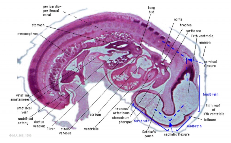
Stomach and lesser sac in mesentery. Triangular flange of mesentery with triangular hole and intervitelline anastomosis. Hindgut (without lumen) seen at caudal end of mesentery.
Neural Development
Musculoskeletal Development
Cervical region: dark masses of dorsal root ganglia. Lumbar region: dorsal aorta with its dorsal segmental arterial branches. Between each dorsal segmental artery is a darker-staining mass of mesenchyme (the dark part of a sclerotome) which is the anlage of the intervertebral disc. The dorsal segmental artery itself marks the location of the centre of the light-staining part of the sclerotome, which is the future vertebral body. The dark band dorsal to the sclerotomes is the basal lamina of the wall of the neural tube.
Original File name: PigG7L.jpg http://embryology.med.unsw.edu.au/wwwpig/pigg/G7L.htm
| System Links: Introduction | Cardiovascular | Coelomic Cavity | Endocrine | Gastrointestinal Tract | Genital | Head | Immune | Integumentary | Musculoskeletal | Neural | Neural Crest | Placenta | Renal | Respiratory | Sensory | Birth |
Cite this page: Hill, M.A. (2024, April 30) Embryology Stage 13 image 098.jpg. Retrieved from https://embryology.med.unsw.edu.au/embryology/index.php/File:Stage_13_image_098.jpg
- © Dr Mark Hill 2024, UNSW Embryology ISBN: 978 0 7334 2609 4 - UNSW CRICOS Provider Code No. 00098G
File history
Click on a date/time to view the file as it appeared at that time.
| Date/Time | Thumbnail | Dimensions | User | Comment | |
|---|---|---|---|---|---|
| current | 17:08, 10 August 2010 |  | 1,000 × 623 (144 KB) | S8600021 (talk | contribs) | ==Stage 13== ==Image Features== ===Gastrointestinal Development=== Stomach and lesser sac in mesentery. Triangular flange of mesentery with triangular hole and intervitelline anastomosis. Hindgut (without lumen) seen at caudal end of mesentery. ===Neur |
You cannot overwrite this file.
File usage
The following 31 pages use this file:
- 2010 Lab 10
- 2010 Lab 5
- 2010 Lab 8
- 2010 Lecture 6
- 2011 Lab 5 - Early Embryo
- ANAT2341 Lab 3 - Week 4
- ANAT2341 Lab 5 - Early Embryo
- ANAT3411 Neuroanatomy
- BGDA Lecture - Development of the Nervous System
- BGDB Gastrointestinal - Early Embryo
- Carnegie stage 13
- Carnegie stage 13 - serial sections
- Embryo Serial Sections
- Fetal ECHO Meeting 2012
- Gastrointestinal Tract - Carnegie Stage 13
- Gastrointestinal Tract - Liver Development
- Lecture - Ectoderm Development
- Lecture - Neural Development
- Museum of Natural History Berlin - 2013 Seminar
- Neural - Mesencephalon Development
- Neural - Prosencephalon Development
- Neural - Rhombencephalon Development
- Neural 3D stage 13 Movie
- Placenta - Stage 13
- RPAH Cardiac Embryology 2014
- Renal System - Carnegie Stage 13
- Respiratory System - Carnegie Stage 13
- Talk:Carnegie stage 13 - serial sections
- Talk:Renal System - Carnegie Stage 13
- Template:Stage13Licon120
- Template talk:Stage13Licon120