File:Stage7 paraxial-mesoderm.jpg: Difference between revisions
No edit summary |
mNo edit summary |
||
| Line 1: | Line 1: | ||
==Carnegie Stage 7 showing the paraxial mesoderm region of the embryonic disc== | ==Carnegie Stage 7 showing the paraxial mesoderm region of the embryonic disc== | ||
{{Stage 7 overlays}} | {{Stage 7 overlays}} | ||
[[Week 3]], [[Carnegie stage 7]], 15 - 17 days, 0.4 mm, embryonic disc, showing the epiblast viewed from the amniotic (dorsal) side. | |||
* Paraxial mesoderm lies lateral to the axial mesoderm. | * Paraxial mesoderm lies lateral to the axial mesoderm. | ||
* During week paraxial mesoderm begins to segment, at the level of the embryo body, into somites. | * During week paraxial mesoderm begins to segment, at the level of the embryo body, into somites. | ||
| Line 8: | Line 10: | ||
* The paraxial mesoderm at the level of the embryo head remains unsegmented. | * The paraxial mesoderm at the level of the embryo head remains unsegmented. | ||
===Carnegie Stage 7=== | |||
Features: embryonic disc, primitive node, primative streak, primative groove, yolk sac | Features: embryonic disc, primitive node, primative streak, primative groove, yolk sac | ||
Revision as of 09:41, 21 June 2013
Carnegie Stage 7 showing the paraxial mesoderm region of the embryonic disc
Stage 7 Mesoderm: axial | paraxial | intermediate | lateral plate
Week 3, Carnegie stage 7, 15 - 17 days, 0.4 mm, embryonic disc, showing the epiblast viewed from the amniotic (dorsal) side.
- Paraxial mesoderm lies lateral to the axial mesoderm.
- During week paraxial mesoderm begins to segment, at the level of the embryo body, into somites.
- historic term was "primitive segments"
- This process is called Somitogenesis
- The paraxial mesoderm at the level of the embryo head remains unsegmented.
Carnegie Stage 7
Features: embryonic disc, primitive node, primative streak, primative groove, yolk sac
Facts: Week 3, 15 - 17 days, 0.4 mm
View 1: embryonic disc, showing the epiblast viewed from the amniotic (dorsal) side.
Events: Gastrulation is continuing as cells migrate from the epiblast, continuing to form mesoderm.
Mesoderm lies between the ectoderm and endoderm as a continuous sheet except at the buccopharyngeal and cloacal membranes. These membranes have ectoderm and endoderm only and will lie at the rostral (head) and caudal (tail) of the gastrointestinal tract.
From the primitive node a tube extends under the ectoderm in the opposite direction to the primitive streak. This tube forms first the axial process then notochordal process, then finally the notochord.
The notochord is a key to embryonic folding and regulation of ectoderm and mesoderm differentiation. It lies in the rostrocordal axis and the embryonic disc will fold either side ventrally, pinching off a portion of the yolk sac to form the lining of the gastrointestinal tract.
- Stage 7 Mesoderm: axial | paraxial | intermediate | lateral plate
Image Source: UNSW Embryology http://embryology.med.unsw.edu.au/wwwhuman/Stages/stage7.htm
No image reuse without permission.
File history
Click on a date/time to view the file as it appeared at that time.
| Date/Time | Thumbnail | Dimensions | User | Comment | |
|---|---|---|---|---|---|
| current | 11:29, 10 August 2009 | 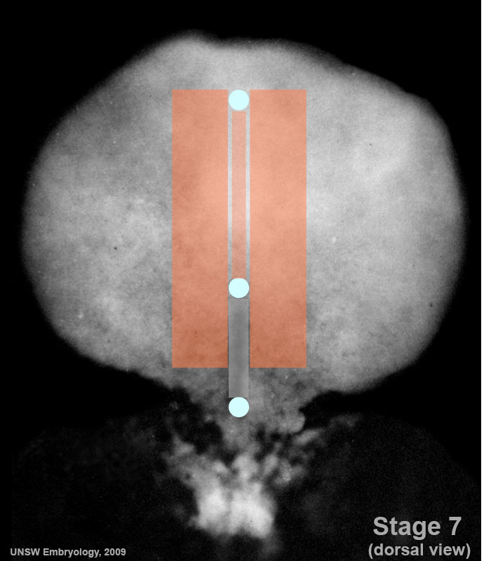 | 690 × 800 (69 KB) | MarkHill (talk | contribs) | Carnegie Stages 7 showing the paraxial mesoderm region of the embryonic disc. Features: embryonic disc, primitive node, primative streak, primative groove, yolk sac Facts: Week 3, 15 - 17 days, 0.4 mm View 1: embryonic disc, showing the epiblast viewed |
You cannot overwrite this file.
File usage
The following 24 pages use this file:
- 2009 Lecture 5
- 2010 BGD Lecture - Development of the Embryo/Fetus 2
- 2010 BGD Practical 6 - Week 3
- 2010 Lab 3
- 2010 Lecture 5
- 2011 Lab 3 - Week 3
- ANAT2341 Lab 3 - Week 3
- BGDA Lecture - Development of the Embryo/Fetus 2
- BGDA Practical 7 - Week 3
- Lecture - Mesoderm Development
- Lecture - Week 3 Development
- Mesoderm
- Talk:2010 BGD Practical 6 - Week 3
- Talk:2011 Lab 3
- File:Stage7-sem2.jpg
- File:Stage7 800x700px.jpg
- File:Stage7 cloacal-oral-membranes.jpg
- File:Stage7 intermediate-mesoderm.jpg
- File:Stage7 lateral-plate.jpg
- File:Stage7 mesoderm.jpg
- File:Stage7 notochord.jpg
- File:Stage7 paraxial-mesoderm.jpg
- File:Stage7 primitive-streak-node.jpg
- Template:Stage 7 mesoderm images