File:Stage7 notochord.jpg: Difference between revisions
From Embryology
mNo edit summary |
|||
| (One intermediate revision by the same user not shown) | |||
| Line 1: | Line 1: | ||
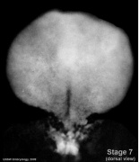
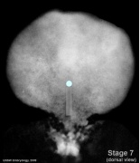
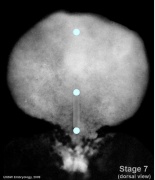
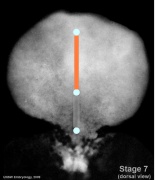
==Carnegie Stage 7 showing the notochord or axial mesoderm region of the embryonic disc== | ==Carnegie Stage 7 showing the notochord or axial mesoderm region of the embryonic disc== | ||
{ | {| | ||
| | |||
* Week 3, 15 - 17 days, 0.4 mm, embryonic disc, showing the epiblast viewed from the amniotic (dorsal) side. | * Week 3, 15 - 17 days, 0.4 mm, embryonic disc, showing the epiblast viewed from the amniotic (dorsal) side. | ||
* Mesoderm lies between the ectoderm and endoderm as a continuous sheet except at the [[B#buccopharyngeal membrane|buccopharyngeal]] and [[C#cloacal membrane|cloacal]] membranes. | * Mesoderm lies between the ectoderm and endoderm as a continuous sheet except at the [[B#buccopharyngeal membrane|buccopharyngeal]] and [[C#cloacal membrane|cloacal]] membranes. | ||
| Line 26: | Line 27: | ||
Events: Gastrulation is continuing as cells migrate from the epiblast, continuing to form mesoderm. | Events: Gastrulation is continuing as cells migrate from the epiblast, continuing to form mesoderm. | ||
| valign=top width=300px| | |||
Image Source: UNSW Embryology | * Blue dot (top) - buccopharyngeal membrane | ||
* Blue dot (middle) - primitive node | |||
* Blue dot (bottom) - cloacal membrane | |||
* Orange line - axial mesoderm (future notochord) | |||
* grey line - primitive streak | |||
:[[Carnegie stage 7|Stage 7 Links]]: :[[Carnegie stage 7]] | [[Gastrulation]] | [[Mesoderm]] | |||
|} | |||
{{Stage 7 mesoderm images}} | |||
===Reference=== | |||
Image Source: UNSW Embryology | |||
No image reuse without permission. | No image reuse without permission. | ||
Latest revision as of 16:04, 17 August 2014
Carnegie Stage 7 showing the notochord or axial mesoderm region of the embryonic disc
Notochord
Features: embryonic disc, primitive node, primative streak, primative groove, yolk sac Facts: Week 3, 15 - 17 days, 0.4 mm View 1: embryonic disc, showing the epiblast viewed from the amniotic (dorsal) side. Events: Gastrulation is continuing as cells migrate from the epiblast, continuing to form mesoderm. |
|
- Embryo Stage 7 (dorsal)
Dorsal view
Primitive streak and node
Oral and cloacal membranes
Axial mesoderm
Paraxial mesoderm
Intermediate mesoderm
Lateral plate


Reference
Image Source: UNSW Embryology
No image reuse without permission.
File history
Click on a date/time to view the file as it appeared at that time.
| Date/Time | Thumbnail | Dimensions | User | Comment | |
|---|---|---|---|---|---|
| current | 11:21, 10 August 2009 | 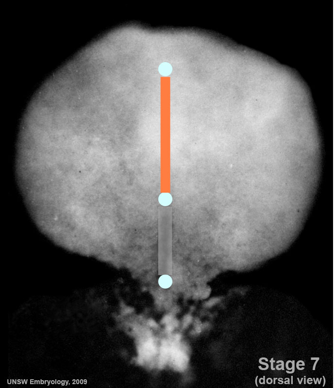 | 690 × 800 (70 KB) | MarkHill (talk | contribs) | Carnegie Stages 7 showing the notochord or axial mesoderm region of the embryonic disc. Features: embryonic disc, primitive node, primative streak, primative groove, yolk sac Facts: Week 3, 15 - 17 days, 0.4 mm View 1: embryonic disc, showing the epibl |
You cannot overwrite this file.
File usage
The following 24 pages use this file:
- 2009 Lecture 5
- 2010 BGD Lecture - Development of the Embryo/Fetus 2
- 2010 BGD Practical 6 - Week 3
- 2010 Lab 3
- 2010 Lecture 5
- 2011 Lab 3 - Week 3
- ANAT2341 Lab 3 - Week 3
- BGDA Lecture - Development of the Embryo/Fetus 2
- BGDA Practical 7 - Week 3
- Lecture - Mesoderm Development
- Lecture - Week 3 Development
- Mesoderm
- Talk:2010 BGD Practical 6 - Week 3
- Talk:2011 Lab 3
- File:Stage7-sem2.jpg
- File:Stage7 800x700px.jpg
- File:Stage7 cloacal-oral-membranes.jpg
- File:Stage7 intermediate-mesoderm.jpg
- File:Stage7 lateral-plate.jpg
- File:Stage7 mesoderm.jpg
- File:Stage7 notochord.jpg
- File:Stage7 paraxial-mesoderm.jpg
- File:Stage7 primitive-streak-node.jpg
- Template:Stage 7 mesoderm images