File:Stage7 bf5a.jpg
Original file (1,024 × 721 pixels, file size: 690 KB, MIME type: image/jpeg)
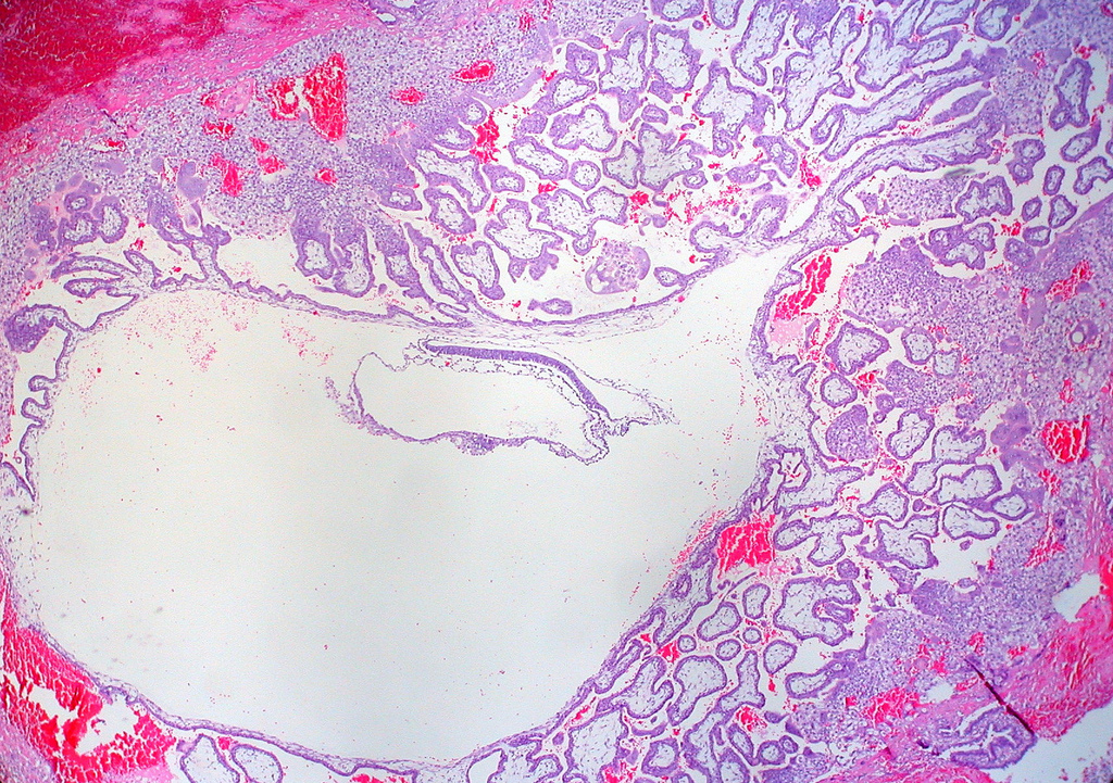
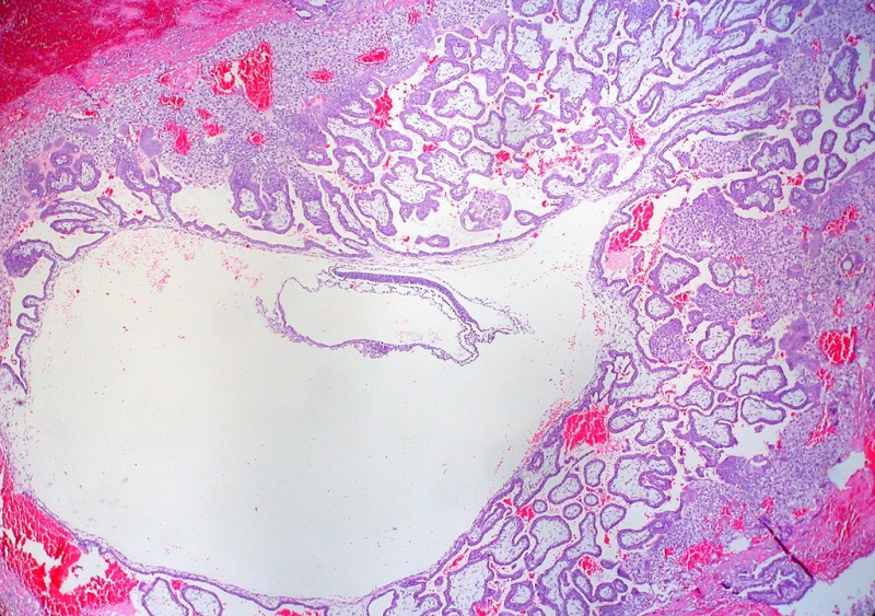
Human embryo - 3 week
Appearance of section looks to be about a Carnegie stage 7 embryo.
Original Author Legend - Primitive Trilaminar Human Embryo in Tubal Pregnancy (40X)
- "I think this is at about the same developmental stage as the Hertig-Rock embryo of 1945, which has been variously estimated at 16.5 or 19 days postovulation (4 to 5 weeks by conventional obstetrical dating, counting from the first day of the last menstrual period)."
Original file name: 3944578509_58800f1ec7_b.jpg
http://www.flickr.com/photos/euthman/3944578509/
Uploaded to Flickr on September 22, 2009 by Ed Uthman Image (pathologist in Houston, Texas)
http://creativecommons.org/licenses/by/2.0/
Image version links: ExtraLarge 1712x1206px | Large 1024x721px | Medium 500x352px | Small 240x169px
Human Embryo Stage 7 Information
Carnegie stage 7
Features: embryonic disc, primitive node, primative streak, primitive groove, yolk sac
Facts: Week 3, 15 - 17 days, 0.4 mm
View 1: embryonic disc, showing the epiblast viewed from the amniotic (dorsal) side.
Events: Gastrulation is continuing as cells migrate from the epiblast, continuing to form mesoderm.
Mesoderm lies between the ectoderm and endoderm as a continuous sheet except at the buccopharyngeal and cloacal membranes. These membranes have ectoderm and endoderm only and will lie at the rostral (head) and caudal (tail) of the gastrointestinal tract.
Original file name: Stage7n3.jpg UNSW Embryology Carnegie Stage 7
Image source: The Kyoto Collection images are reproduced with the permission of Prof. Kohei Shiota and Prof. Shigehito Yamada, Anatomy and Developmental Biology, Kyoto University Graduate School of Medicine, Kyoto, Japan for educational purposes only and cannot be reproduced electronically or in writing without permission.
File history
Click on a date/time to view the file as it appeared at that time.
| Date/Time | Thumbnail | Dimensions | User | Comment | |
|---|---|---|---|---|---|
| current | 14:44, 21 July 2010 | | 1,024 × 721 (690 KB) | S8600021 (talk | contribs) | ==Human embryo - 3 week== Appearance of section looks to be about a Carnegie stage 7 embryo. Original Author Legend - Primitive Trilaminar Human Embryo in Tubal Pregnancy (40X) :"I think this is at about the same developmental stage as the Hertig-Roc |
You cannot overwrite this file.
File usage
There are no pages that use this file.