File:Stage11 sem6.jpg
Original file (612 × 1,000 pixels, file size: 57 KB, MIME type: image/jpeg)
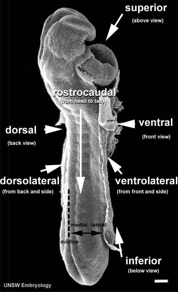
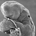
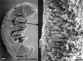
Human Embryo Carnegie stage 11
This scanning EM of the stage 11 human embryo is the same image as Stage11 sem5 with the addition of direction of view.
Alternative Terms
- ventral and anterior both mean toward the front of the body.
- dorsal and posterior mean the back of the body.
- rostral, cranial, and cephalic and superior all mean toward the head or the upper part of a structure
- proximal means closer to the body
- distal is away from the body
- medial describes a structure toward the midline of the body
- lateral describes a structure away from the midline of the body
Body Planes
- coronal or frontal plane - divides the body into ventral and dorsal
- sagittal - divides the body into left and right
- midsagittal or median sagittal - is the plane exactly in the midline
- transverse or horizontal plane - divides the body into rostral and caudal
About this Embryo
Carnegie stage 11 24 days, 13 somite pairs
Facts: Week 4, 23 - 26 days, 2.5 - 4.5 mm, Somite Number 13 - 20
View: This is a scanning EM of the embryo dorsal view showing the neural tube closing with open neuropores and the paired somites visible through the thin ectoderm.
Features: surface ectoderm, neural tube, cranial (anterior) neuropore, caudal (posterior) neuropore, somites, heart, cut edge of amnion
Stage11_sem6.jpg
Original file name: Stage11day24somite13-dorsal-sem5-1000px.jpg
- Stage 11 SEM Images: dorsolateral whole embryo | dorsal embryo | lateral embryo | lateral head | lateral head with overlay | embryo cross-section | ventrolateral head | ventrolateral head with overlay | ventral head | buccopharyngeal membrane | neural crest | posterior neuropore | anterior neuropore | Carnegie stage 11
- Human Embryo (stage 11)
dorsolateral whole embryo
dorsal embryo
lateral embryo
lateral head
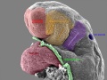
lateral head with overlay

embryo cross-section
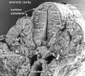
embryo cross-section label
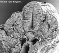
neural cross-section label

ventrolateral head
ventrolateral head with overlay

ventral head

buccopharyngeal membrane
neural crest
posterior neuropore
anterior neuropore


Image Source: Scanning electron micrographs of the Carnegie stages of the early human embryos are reproduced with the permission of Prof Kathy Sulik, from embryos collected by Dr. Vekemans and Tania Attié-Bitach. Images are for educational purposes only and cannot be reproduced electronically or in writing without permission.
File history
Click on a date/time to view the file as it appeared at that time.
| Date/Time | Thumbnail | Dimensions | User | Comment | |
|---|---|---|---|---|---|
| current | 14:13, 5 May 2011 | | 612 × 1,000 (57 KB) | S8600021 (talk | contribs) | |
| 13:49, 5 May 2011 | 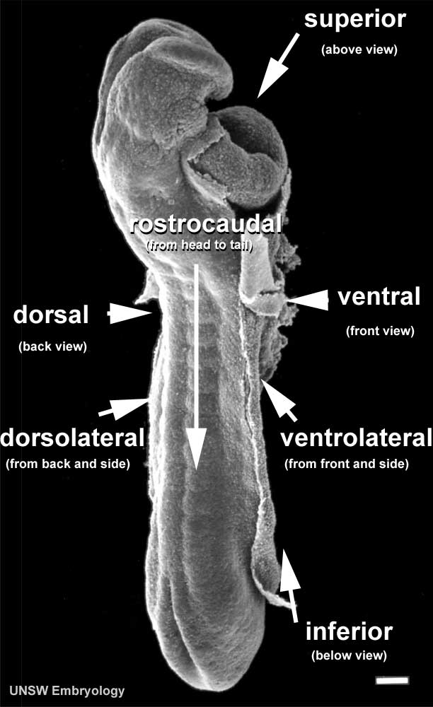 | 612 × 1,000 (57 KB) | S8600021 (talk | contribs) | ==Human Embryo Carnegie stage 11== This scanning EM of the stage 11 human embryo is the same image as Stage11 sem5 with the addition of Carnegie stage 11 24 days, 13 somite pairs Facts: Week 4, 23 - 26 days, 2.5 - 4.5 mm, So |
You cannot overwrite this file.
File usage
The following 21 pages use this file:
- 2009 Lecture 6
- 2010 BGD Lecture - Development of the Embryo/Fetus 1
- 2011 Lab 3 - Week 4
- ANAT2341 Lab 3 - Week 4
- Abnormal Development - Thalidomide
- BGDA Lecture - Development of the Embryo/Fetus 1
- BGDA Lecture - Development of the Embryo/Fetus 2
- BGDA Lecture - Development of the Nervous System
- BGDA Practical - Implantation to 8 Weeks
- BGDA Practical 7 - Week 4
- Carnegie stage 11
- Developmental Mechanism - Axes Formation
- Developmental Mechanism - Dorso-Ventral Axis
- Developmental Mechanism - Left-Right Axis
- Developmental Mechanism - Rostro-Caudal axis Axis
- Human Embryo - Scanning electron microscopy
- Human Embryo SEM
- K12 Thalidomide
- Talk:2011 Lab 3
- Talk:Carnegie stage 11
- Template:Carnegie stage 11-14 image table