File:Spleen anatomy.jpg: Difference between revisions
From Embryology
m (→Spleen Anatomy) |
|||
| Line 1: | Line 1: | ||
==Spleen Anatomy== | ==Spleen Anatomy== | ||
* The spleen is located in the upper left abdominal cavity, just beneath the diaphragm, and posterior to the stomach. | * The {{spleen}} is located in the upper left abdominal cavity, just beneath the diaphragm, and posterior to the stomach. | ||
* It is similar to a lymph node in shape and structure but it is much larger. | * It is similar to a lymph node in shape and structure but it is much larger. | ||
* The spleen is the largest lymphatic organ in the body. | * The spleen is the largest lymphatic organ in the body. | ||
| Line 11: | Line 11: | ||
* Blood enters the spleen through the splenic artery, moves through the sinuses where it is filtered, then leaves through the splenic vein. | * Blood enters the spleen through the splenic artery, moves through the sinuses where it is filtered, then leaves through the splenic vein. | ||
{{Footer}} | |||
==Function== | ==Function== | ||
Revision as of 12:05, 26 July 2019
Spleen Anatomy
- The spleen is located in the upper left abdominal cavity, just beneath the diaphragm, and posterior to the stomach.
- It is similar to a lymph node in shape and structure but it is much larger.
- The spleen is the largest lymphatic organ in the body.
- Surrounded by a connective tissue capsule, which extends inward to divide the organ into lobules, the spleen consists of two types of tissue called white pulp and red pulp.
White pulp - is lymphatic tissue consisting mainly of lymphocytes around arteries.
Red pulp - consists of venous sinuses filled with blood and cords of lymphatic cells, such as lymphocytes and macrophages.
- Blood enters the spleen through the splenic artery, moves through the sinuses where it is filtered, then leaves through the splenic vein.
Cite this page: Hill, M.A. (2024, May 3) Embryology Spleen anatomy.jpg. Retrieved from https://embryology.med.unsw.edu.au/embryology/index.php/File:Spleen_anatomy.jpg
- © Dr Mark Hill 2024, UNSW Embryology ISBN: 978 0 7334 2609 4 - UNSW CRICOS Provider Code No. 00098G
Function
Immune
- filters blood in much the way that the lymph nodes filter lymph.
- Lymphocytes in the spleen react to pathogens in the blood and attempt to destroy them.
- Macrophages then engulf the resulting debris, the damaged cells, and the other large particles.
Red Blood Cell Removal
- The spleen (and liver) removes old and damaged erythrocytes from the circulating blood.
- Like other lymphatic tissue, it produces lymphocytes, especially in response to invading pathogens.
Blood Reservoir
- The sinuses in the spleen also act as a reservoir for blood.
- In emergencies, such as hemorrhage, smooth muscle in the vessel walls and in the capsule of the spleen contracts.
- This squeezes the blood out of the spleen into the general circulation.
Image and Modified text source: National Cancer Institute - SEER Training Modules
http://training.seer.cancer.gov/anatomy/lymphatic/components/spleen.html
http://training.seer.cancer.gov/anatomy/lymphatic/components/
File history
Click on a date/time to view the file as it appeared at that time.
| Date/Time | Thumbnail | Dimensions | User | Comment | |
|---|---|---|---|---|---|
| current | 14:27, 21 February 2011 | 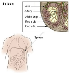 | 275 × 285 (19 KB) | S8600021 (talk | contribs) |
You cannot overwrite this file.
File usage
The following 3 pages use this file: