File:Small intestine secondary loops week 7to8.jpg
Original file (1,280 × 837 pixels, file size: 168 KB, MIME type: image/jpeg)
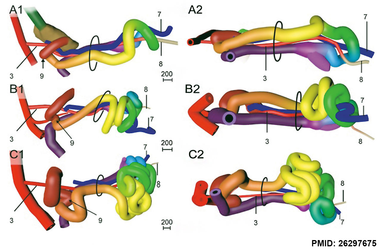
Small intestine secondary loops week 7 and 8
Right-lateral (panels a1-c1) and caudal (panels a2-c2) views of the reconstructed intestines of CS18 (s97; panel a), CS20 (462; panel b), and CS23 (s4141; panel c) embryos.
The beige strand (8) represents the periductal mesenchyme. Ovals: umbilical orifice. Note the dorsal (a) and then leftward growth (b, c) of the apex of the 1st secondary loop (coded red and orange), followed by the formation of tertiary loops in its distal limb (c; coded orange). Further note the caudal (a) and then leftward growth (b, c) of the apex of the 2nd and 3rd secondary loops (coded yellow and green, respectively), followed by the formation of tertiary loops (c). Also note that the apex of the 4th secondary loop (coded blue) grows cranially (a) before forming tertiary loops (c).
Scale bar units: μm. An interactive 3D-PDF is available online (3D-PDF CS20)
- Links: week 5 | secondary loops week 7 to 8 | tertiary loops week 8 | Intestine Development | Week 7 | Week 8
Reference
Soffers JH, Hikspoors JP, Mekonen HK, Koehler SE & Lamers WH. (2015). The growth pattern of the human intestine and its mesentery. BMC Dev. Biol. , 15, 31. PMID: 26297675 DOI.
Copyright
© 2015 Soffers et al. Open Access This article is distributed under the terms of the Creative Commons Attribution 4.0 International License (http://creativecommons.org/licenses/by/4.0), which permits unrestricted use, distribution, and reproduction in any medium, provided you give appropriate credit to the original author(s) and the source, provide a link to the Creative Commons license, and indicate if changes were made. The Creative Commons Public Domain Dedication waiver (http://creativecommons.org/publicdomain/zero/1.0/) applies to the data made available in this article, unless otherwise stated.
Fig. 4 Image resized and PMID added.
Cite this page: Hill, M.A. (2024, April 27) Embryology Small intestine secondary loops week 7to8.jpg. Retrieved from https://embryology.med.unsw.edu.au/embryology/index.php/File:Small_intestine_secondary_loops_week_7to8.jpg
- © Dr Mark Hill 2024, UNSW Embryology ISBN: 978 0 7334 2609 4 - UNSW CRICOS Provider Code No. 00098G
File history
Click on a date/time to view the file as it appeared at that time.
| Date/Time | Thumbnail | Dimensions | User | Comment | |
|---|---|---|---|---|---|
| current | 12:51, 24 January 2017 | | 1,280 × 837 (168 KB) | Z8600021 (talk | contribs) | |
| 12:51, 24 January 2017 |  | 1,989 × 1,216 (346 KB) | Z8600021 (talk | contribs) | Fig. 4 Development of secondary loops in the small intestine during the 7th and 8th week is invariant. Right-lateral (panels a1-c1) and caudal (panels a2-c2) views of the reconstructed intestines of CS18 (s97; panel a), CS20 (462; panel b), and CS23 (s... |
You cannot overwrite this file.
File usage
The following 2 pages use this file: