File:Skeletal muscle histology 016.jpg: Difference between revisions
(==Human Skeletal Muscle Histology== * Human skeletal muscle * longitudinal section of muscle fibres * sarcomeres visible * Stain HE magnification x40 * See also full image {{SkMhistolinks}} {{Blue Histology}) |
No edit summary |
||
| Line 1: | Line 1: | ||
==Human Skeletal Muscle Histology== | ==Human Skeletal Muscle Histology== | ||
Human skeletal muscle longitudinal section of muscle fibres showing sarcomeres. Stain HE magnification x40. See also [[:File:Skeletal_muscle_histology_013.jpg|full image]] | |||
Sarcomere components | |||
* '''A band''' - (anisotropic bands, light band) are composed of the thick myosin filaments. | |||
* '''I band''' - (isotropic bands, light band) are composed of thin actin filaments. | |||
* '''H band''' - (German, ''heller'' =brighter). are composed of the thick filaments that is not overlapped (superimposed) by the thin filaments. | |||
* '''Z line''' - (German, ''Zwischenscheibe'' = Intermediate plate) appears as a series of dark lines between the I bands, indicate the end of one sarcomere and the beginning of the next. | |||
:'''Links:''' [[:File:Sarcomere animation.gif|Sarcomere animation]] | [[:File:Skeletal_muscle_histology_016.jpg|Sarcomere histology]] | [[Skeletal_Muscle_Histology|Muscle Histology]] | [[Musculoskeletal_System_-_Muscle_Development|Muscle Development]] | |||
|} | |||
{{SkMhistolinks}} | {{SkMhistolinks}} | ||
Revision as of 10:27, 6 March 2012
Human Skeletal Muscle Histology
Human skeletal muscle longitudinal section of muscle fibres showing sarcomeres. Stain HE magnification x40. See also full image
Sarcomere components
- A band - (anisotropic bands, light band) are composed of the thick myosin filaments.
- I band - (isotropic bands, light band) are composed of thin actin filaments.
- H band - (German, heller =brighter). are composed of the thick filaments that is not overlapped (superimposed) by the thin filaments.
- Z line - (German, Zwischenscheibe = Intermediate plate) appears as a series of dark lines between the I bands, indicate the end of one sarcomere and the beginning of the next.
|}
- Muscle Histology: Muscle Development | Human HE x4 longitudinal and transverse | Human HE x40 transverse | Human HE x40 longitudinal | Human HE x40 longitudinal | Human HE x4 longitudinal and transverse | Muscle Spindle HE x40 | Human HE x40 | Human HE x40 | Human HE x40 | Human HE x100 | Human HE x100 | Fetal human muscle | Myotendinous junction label | Myotendinous junction HE x40 | Whipf 1 | Whipf 2 | Whipf 3 | Tongue HE x10 transverse | Tongue x100 | Muscle spindle HE x20 | Muscle spindle HE x40
Links: Histology | Histology Stains | Blue Histology images copyright Lutz Slomianka 1998-2009. The literary and artistic works on the original Blue Histology website may be reproduced, adapted, published and distributed for non-commercial purposes. See also the page Histology Stains.
Cite this page: Hill, M.A. (2024, May 19) Embryology Skeletal muscle histology 016.jpg. Retrieved from https://embryology.med.unsw.edu.au/embryology/index.php/File:Skeletal_muscle_histology_016.jpg
- © Dr Mark Hill 2024, UNSW Embryology ISBN: 978 0 7334 2609 4 - UNSW CRICOS Provider Code No. 00098G
Skem040he2.jpg
File history
Click on a date/time to view the file as it appeared at that time.
| Date/Time | Thumbnail | Dimensions | User | Comment | |
|---|---|---|---|---|---|
| current | 01:31, 3 October 2011 | 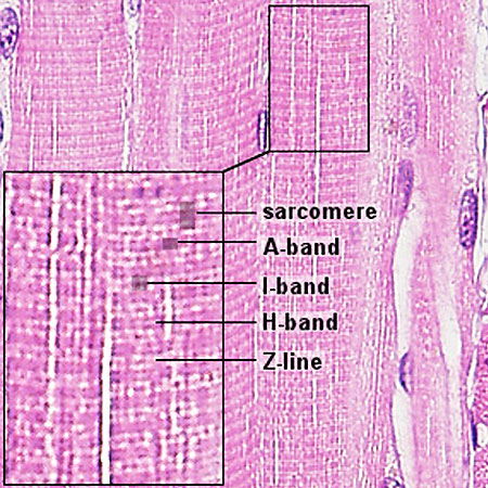 | 450 × 450 (92 KB) | S8600021 (talk | contribs) | ==Human Skeletal Muscle Histology== * Human skeletal muscle * longitudinal section of muscle fibres * sarcomeres visible * Stain HE magnification x40 * See also full image {{SkMhistolinks}} {{Blue Histology} |
You cannot overwrite this file.