File:Sheep follicle in vitro 01.jpg: Difference between revisions
| (One intermediate revision by the same user not shown) | |||
| Line 5: | Line 5: | ||
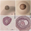
On the left panels, examples of preantral (PA) follicles used for the culture and analyzed under a stereomicroscope (on the top) and after staining with haematoxylin/eosin (HE) (at the bottom). On the right panels, early antral (EA) follicles cultured for 14 days and analyzed under a stereomicroscope (on the top) and after staining with haematoxylin/eosin (at the bottom). Either in PA and EA follicles HE staining showed the presence of a compact layers of granulosa cells and a theca compartment with several fusiform cells (arrows). | On the left panels, examples of preantral (PA) follicles used for the culture and analyzed under a stereomicroscope (on the top) and after staining with haematoxylin/eosin (HE) (at the bottom). On the right panels, early antral (EA) follicles cultured for 14 days and analyzed under a stereomicroscope (on the top) and after staining with haematoxylin/eosin (at the bottom). Either in PA and EA follicles HE staining showed the presence of a compact layers of granulosa cells and a theca compartment with several fusiform cells (arrows). | ||
:'''Links:''' [[Sheep Development]] | [[Oocyte Development]] | |||
===Reference=== | ===Reference=== | ||
| Line 10: | Line 14: | ||
Copyright | ====Copyright==== | ||
© 2011 Barboni et al. This is an open-access article distributed under the terms of the Creative Commons Attribution License, which permits unrestricted use, distribution, and reproduction in any medium, provided the original author and source are credited. | |||
Latest revision as of 21:51, 6 June 2013
Sheep Follicle in vitro
Representative images of the morphology of the follicles before and after the culture.
On the left panels, examples of preantral (PA) follicles used for the culture and analyzed under a stereomicroscope (on the top) and after staining with haematoxylin/eosin (HE) (at the bottom). On the right panels, early antral (EA) follicles cultured for 14 days and analyzed under a stereomicroscope (on the top) and after staining with haematoxylin/eosin (at the bottom). Either in PA and EA follicles HE staining showed the presence of a compact layers of granulosa cells and a theca compartment with several fusiform cells (arrows).
- Links: Sheep Development | Oocyte Development
Reference
<pubmed>22132111</pubmed>| PLoS One.
Copyright
© 2011 Barboni et al. This is an open-access article distributed under the terms of the Creative Commons Attribution License, which permits unrestricted use, distribution, and reproduction in any medium, provided the original author and source are credited.
Figure 1. doi:info:doi/10.1371/journal.pone.0027550.g001
Journal.pone.0027550.g001.png
converted to jpg
File history
Click on a date/time to view the file as it appeared at that time.
| Date/Time | Thumbnail | Dimensions | User | Comment | |
|---|---|---|---|---|---|
| current | 01:39, 7 June 2012 |  | 600 × 600 (108 KB) | Z8600021 (talk | contribs) | ==Sheep Follicle in vitro== Representative images of the morphology of the follicles before and after the culture. On the left panels, examples of preantral (PA) follicles used for the culture and analyzed under a stereomicroscope (on the top) and afte |
You cannot overwrite this file.
File usage
The following page uses this file: