File:Reid1908 fig01.jpg: Difference between revisions
(===Reference=== {{Ref-Reid1908}}) |
mNo edit summary |
||
| Line 1: | Line 1: | ||
==Fig. 1. == | |||
The surfaces of the duodenum, jejunum, and pancreas, which were in apposition with each other, were bare as regards peritoneum. The posterior aspect of the third portion of the duodenum was free behind, the peritoneum leaving the upper border of this aspect to reach the posterior abdominal wall along an almost horizontal line which formed the upper duodenal portion of the line of soldering (see fig., D) to be described. Inasmuch as the second part of the duodenum, head of the pancreas, and jejunum came into antero-posterior relation with one another the mesentery of the last named was common to all. All were thus equally mobile. Above the jejunum a lamina of peritoneum (part of the originally right lamina of the mesoduodenum), lying in the frontal plane, and about 2 cms. long, connected the second part of the duodenum to the posterior abdominal wall quite to the right of the superior mesenteric artery. Upon the front of this lamina was a rather well-developed muscle of Treitz embedded in connective tissue, representing part of the originally left lamina of the mesoduodenum. | |||
===Reference=== | ===Reference=== | ||
{{Ref-Reid1908}} | {{Ref-Reid1908}} | ||
Revision as of 18:57, 19 March 2018
Fig. 1.
The surfaces of the duodenum, jejunum, and pancreas, which were in apposition with each other, were bare as regards peritoneum. The posterior aspect of the third portion of the duodenum was free behind, the peritoneum leaving the upper border of this aspect to reach the posterior abdominal wall along an almost horizontal line which formed the upper duodenal portion of the line of soldering (see fig., D) to be described. Inasmuch as the second part of the duodenum, head of the pancreas, and jejunum came into antero-posterior relation with one another the mesentery of the last named was common to all. All were thus equally mobile. Above the jejunum a lamina of peritoneum (part of the originally right lamina of the mesoduodenum), lying in the frontal plane, and about 2 cms. long, connected the second part of the duodenum to the posterior abdominal wall quite to the right of the superior mesenteric artery. Upon the front of this lamina was a rather well-developed muscle of Treitz embedded in connective tissue, representing part of the originally left lamina of the mesoduodenum.
Reference
Reid DG. Imperfect torsion of the intestinal loop. (1908) J Anat Physiol. 42(3): 320-305. PMID 17232774
File history
Click on a date/time to view the file as it appeared at that time.
| Date/Time | Thumbnail | Dimensions | User | Comment | |
|---|---|---|---|---|---|
| current | 18:57, 19 March 2018 | 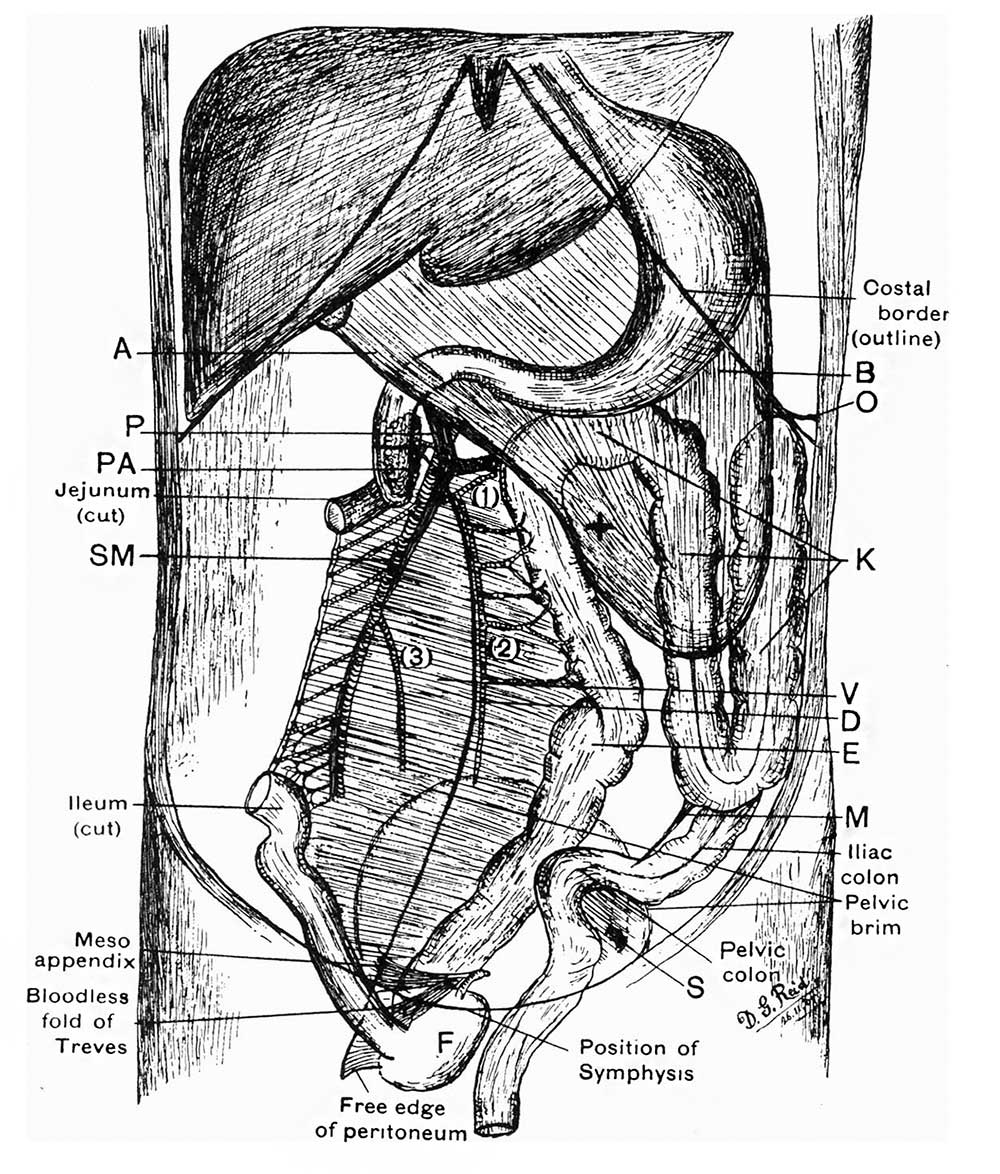 | 1,000 × 1,174 (252 KB) | Z8600021 (talk | contribs) | |
| 18:54, 19 March 2018 | 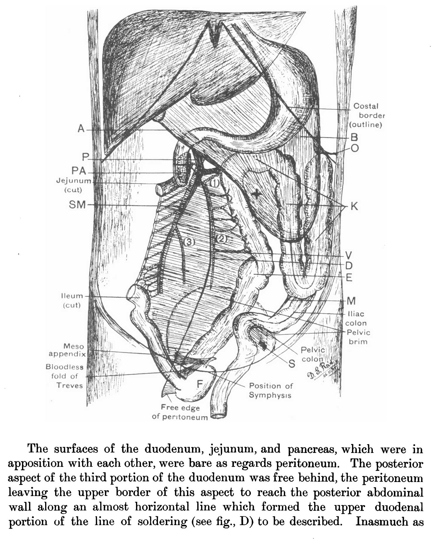 | 1,474 × 1,851 (353 KB) | Z8600021 (talk | contribs) | ===Reference=== {{Ref-Reid1908}} |
You cannot overwrite this file.
File usage
The following page uses this file: