File:Rat neural cadherin 08.jpg

Original file (597 × 678 pixels, file size: 31 KB, MIME type: image/jpeg)
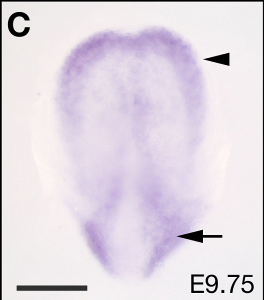
Expression patterns of cad7 in the developing rat E9.75 embryo
A-C: The expression of cad7 mRNA in the anterior margin of the early neural plate at E9.5–9.75 (arrowhead in A, B and C), and in the lateral plate at E9.75 (arrow in C). These pictures show lateral (A), ventral (B) and dorsal (C) views. D: Lateral view showing the expression of cad7 in the brain region anterior to the midbrain/hindbrain boundary (arrowhead) and caudal neural tube (arrow) at E10.5. D': Dorsal view of D. The expression of cad7 is detected at the edge of the neural plate and part of migrating neural crest cells (arrowheads). E-G: Expression of cad7 in the forebrain and midbrain (E). Cad7 is expressed in the dorsal region of the otic vesicle (ov), and in the olfactory epithelium (oe) and retina (r) at E11.5 (E). G is a cross-section at the fore limb (fl) level. In the hindbrain and spinal cord, cad7 is expressed in the dorsal neuroepithelium and the expression is absent in the roof plate (rp) (F, G). E and F images indicate lateral and ventral view, respectively. H: Lateral view of cad7 expression in the pharyngeal region at E12.0. Arrow and arrowhead indicate expression of cad7 in the pharyngeal groove (pg) and otic vesicle (ov), respectively. I-J: Lateral view of cad7 staining in the brain of E12.5 embryos. Arrow in I indicates the expression of cad7 in the ventral domains of prosomere3 (p3) and secondary prosencephalon. No expression of cad7 in the dorsal root ganglion cells at the trunk level (J). K-N: On cross-sections from E14.5 embryo, cad7 transcripts are detected in the subpopulation of motor neurons (mn) but not in the dorsal root ganglion or Schwann cell precursors (arrowheads in K, and M) expressing Sox10, along motor nerve (arrowheads in L and N). al, pharyngeal arch 1; da, dorsal aorta; ctx, cerebral cortex; mb, midbrain. Scale bars: 200 μm in A-C, G and K-N; 400 μm in D; 100 μm in D'; 500 μm in E and H; 300 μm in F; 1 mm in I and J.
Reference
<pubmed>18801203</pubmed>| PMC2564927 | BMC Dev Biol.
© 2008 Takahashi and Osumi; licensee BioMed Central Ltd.
This is an Open Access article distributed under the terms of the Creative Commons Attribution License (http://creativecommons.org/licenses/by/2.0), which permits unrestricted use, distribution, and reproduction in any medium, provided the original work is properly cited.
1471-213X-8-87-3.jpg
1471-213X-8-87-2.jpg
File history
Click on a date/time to view the file as it appeared at that time.
| Date/Time | Thumbnail | Dimensions | User | Comment | |
|---|---|---|---|---|---|
| current | 19:00, 23 June 2012 | | 597 × 678 (31 KB) | Z8600021 (talk | contribs) | ==Expression patterns of cad7 in the developing rat E9.75 embryo== A-C: The expression of cad7 mRNA in the anterior margin of the early neural plate at E9.5–9.75 (arrowhead in A, B and C), and in the lateral plate at E9.75 (arrow in C). These pictures |
You cannot overwrite this file.
File usage
The following page uses this file: