File:Pohlman1911 plate2D.jpg
Original file (1,822 × 1,397 pixels, file size: 166 KB, MIME type: image/jpeg)
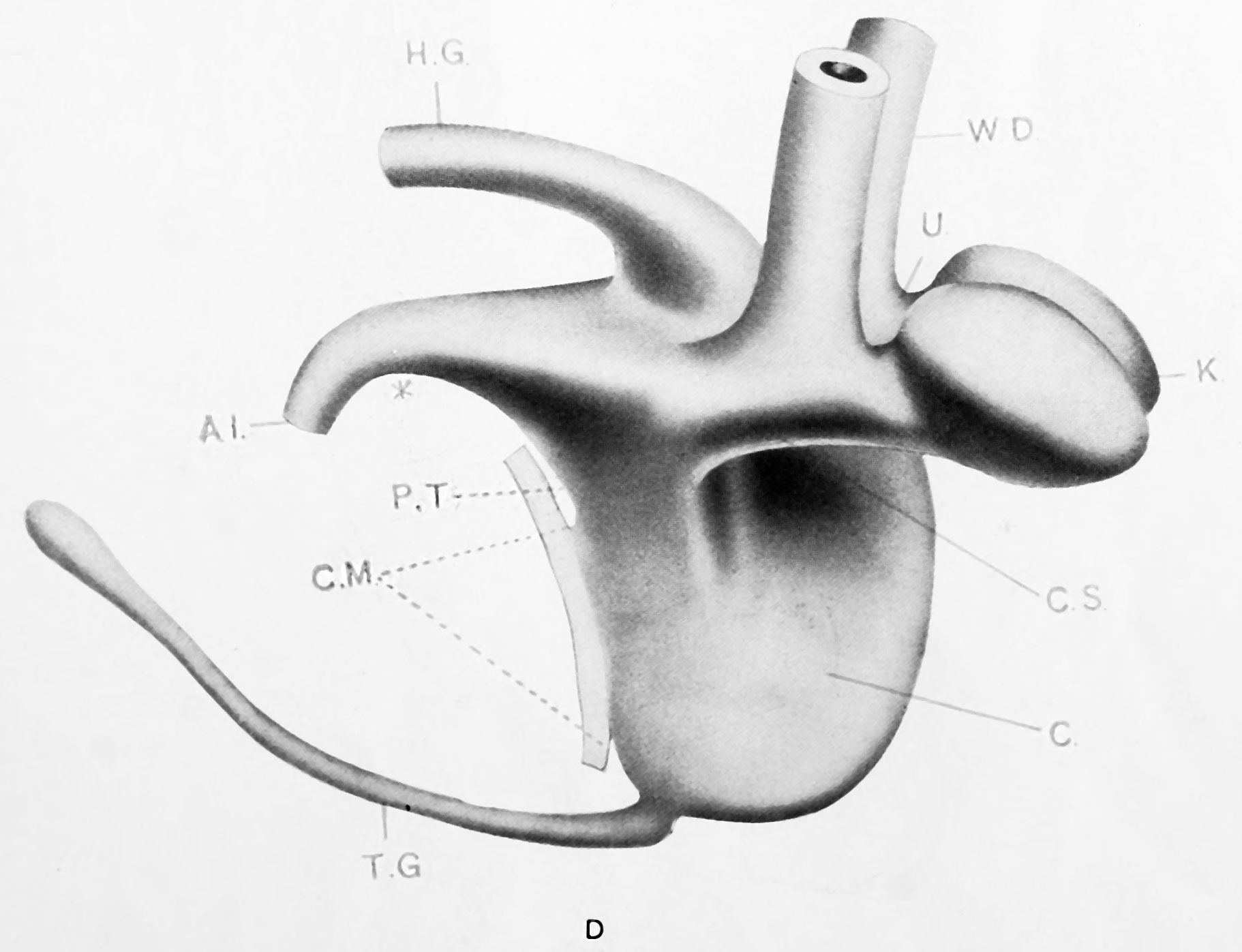
Plate 2D
No. 12 (Mall 2; 7.0 mm.; +; 15; Good). Model D x 100. Marked development of renal buds. Kidney and ureter segments evident. Ventral cloacal segment much widened. Allantois of narrow lumen. Intracloacal epithelial plug marked. Tail gut maximum in length and slightly widened at caudal end. Carnegie stage 15
Model D {No. 12; Mall no. 2; 7.0 mm.) This model fills in a gap in the Keibel series which may be considered quite an important one. The hind gut and all that pertains to the dorsal segment of the cloaca has retained an even caliber, while the ventral segment has widened progressively. The division of the cloaca may be traced to the level of the Wolffian orifices. The Wolffian ducts are much better developed and the renal anlage has resolved itself into distinct ureter and kidney segments. The segment of Wolffian duct between the orifice of the ureter and the cloaca is relatively shorter. The renal anlagen have assumed the position of dorsal convergence (mentioned by Keibel) but this is only a relative matter (see later). The tail gut has undergone further degeneration and has lost its lumen in part.
| Key to Lettering | ||
|---|---|---|
|
A., Allantois K., Kidney A.M., Position of anal membrane P.T., Precloacal mesodermic tissue C., Cloaca |
R.B., Renal bud CM., Cloacal membrane T.G., Tail cut C.S., Cloacal segment of Wolffian duct U., Ureter |
G.E., Genital eminence U.G., Urogenital sinus H.G., Hind gut U.M., Position of urogenital membrane * Probable position of point where allantois joins cloaca. |
All drawings represent 100 diameters enlargement except of Model F which is 50 diameters.
- Links: plate 1 | 1 A | 1 B | plate 2 | plate 2C | plate 2D | plate 3 | plate 3E | plate 3F | Pohlman 1911
Reference
Pohlman AG. The development of the cloaca in human embryos. (1911) Amer. J Anat. 12: 1-26.
Cite this page: Hill, M.A. (2024, April 28) Embryology Pohlman1911 plate2D.jpg. Retrieved from https://embryology.med.unsw.edu.au/embryology/index.php/File:Pohlman1911_plate2D.jpg
- © Dr Mark Hill 2024, UNSW Embryology ISBN: 978 0 7334 2609 4 - UNSW CRICOS Provider Code No. 00098G
File history
Click on a date/time to view the file as it appeared at that time.
| Date/Time | Thumbnail | Dimensions | User | Comment | |
|---|---|---|---|---|---|
| current | 14:41, 15 June 2016 | | 1,822 × 1,397 (166 KB) | Z8600021 (talk | contribs) | ==Plate 2D== {{Pohlman1911 figures}} |
You cannot overwrite this file.
File usage
The following page uses this file: