File:Pohlman1911 fig1.jpg

Original file (769 × 675 pixels, file size: 33 KB, MIME type: image/jpeg)
Fig. 1.
An ectodermal cloaca has been stated by Keibel to be present only in traces, if at all. None of the embryos examined showed a surface depression, and we may therefore critically examine Nagel's fig. 1 and his contention concerning the cloacal membrane. It will be seen that in fig. 1, while the embryo shows a cloacal division comparable to our Model E; the phallus is developed to the extent of that found in Model F ; while the cloacal membrane is ruptured. None of the embryos of this stage, or younger than this stage, showed rupture of the cloacal membrane, and we may therefore agree with Keibel that the cloacal membrane remains intact normally as long as it persists as such. Nagel believes that the rupture of the membrane is essential or where would the secretion of the mesonephros be passed? We may answer this question. The ventral cloacal segment and the allantois do not show the degree of widening necessary to accommodate the urinary excretion, and we call to mind Hill's work on the pig embryo ('04) and our own ('04) on the human embryo, in both of which it is conclusively shown that the mesonephros is actively degenerate long before the kidney is developmentally fitted to take up a urinary function. Cases of atresia urethrae commonly occur without bladder distension ; monstrosities may go to term without kidneys and with no undue persistence of the mesonephros; and experimentally, it has been shown that there is no evidence of urinary excretion into the amniotic fluid even in term children. We do not believe it necessary to assume that the mesonephros functionates as an organ of excretion in the placental mammals, and do not consider Nagel's contention for a functional rupture of the cloacal membrane as a serious one.
| Key to Lettering | ||
|---|---|---|
|
A., Allantois K., Kidney A.M., Position of anal membrane P.T., Precloacal mesodermic tissue C., Cloaca |
R.B., Renal bud CM., Cloacal membrane T.G., Tail cut C.S., Cloacal segment of Wolffian duct U., Ureter |
G.E., Genital eminence U.G., Urogenital sinus H.G., Hind gut U.M., Position of urogenital membrane * Probable position of point where allantois joins cloaca. |
All drawings represent 100 diameters enlargement except of Model F which is 50 diameters.
- Links: plate 1 | 1 A | 1 B | plate 2 | plate 2C | plate 2D | plate 3 | plate 3E | plate 3F | Pohlman 1911
Reference
Pohlman AG. The development of the cloaca in human embryos. (1911) Amer. J Anat. 12: 1-26.
Cite this page: Hill, M.A. (2024, April 27) Embryology Pohlman1911 fig1.jpg. Retrieved from https://embryology.med.unsw.edu.au/embryology/index.php/File:Pohlman1911_fig1.jpg
- © Dr Mark Hill 2024, UNSW Embryology ISBN: 978 0 7334 2609 4 - UNSW CRICOS Provider Code No. 00098G
File history
Click on a date/time to view the file as it appeared at that time.
| Date/Time | Thumbnail | Dimensions | User | Comment | |
|---|---|---|---|---|---|
| current | 11:52, 12 June 2016 | | 769 × 675 (33 KB) | Z8600021 (talk | contribs) | |
| 11:51, 12 June 2016 | 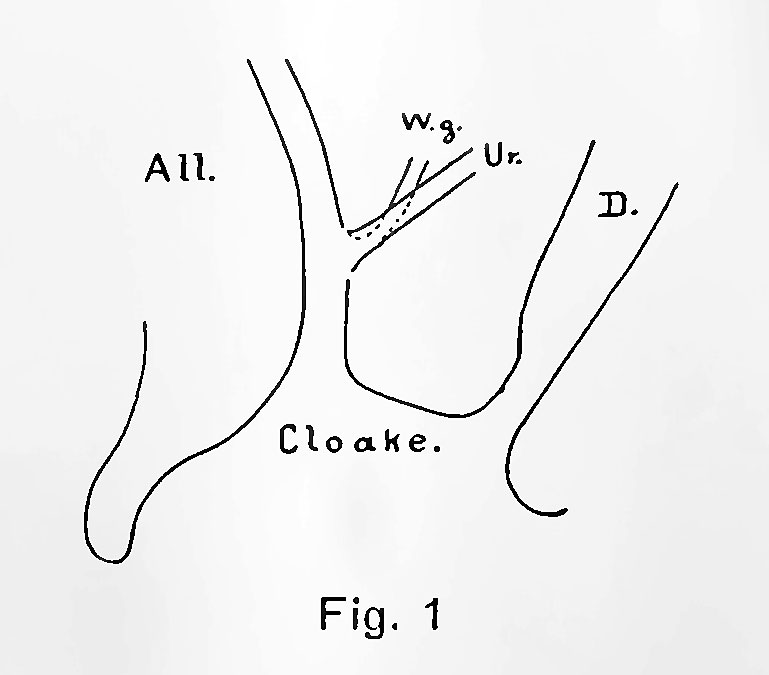 | 769 × 675 (37 KB) | Z8600021 (talk | contribs) | {{Pohlman1911 Figures}} |
You cannot overwrite this file.
File usage
The following page uses this file: