File:Placenta MRI 01.jpg: Difference between revisions
mNo edit summary |
mNo edit summary |
||
| Line 5: | Line 5: | ||
:[[Magnetic Resonance Imaging|'''MRI Links''']]: [[:File:Placenta MRI 02.jpg|image - placenta 22 and 32wk]] | [[:File:Placenta MRI 01.jpg|image - placenta 37wk]] | [[Placenta Development]] | [[Magnetic Resonance Imaging]] | |||
===Reference=== | ===Reference=== | ||
<pubmed>24604945</pubmed>| [http://www.ncbi.nlm.nih.gov/pmc/articles/PMC3932583 PMC3932583] | [http://www.ijri.org/text.asp?2013/23/4/379/125592 Indian J Radiol Imaging.] | <pubmed>24604945</pubmed>| [http://www.ncbi.nlm.nih.gov/pmc/articles/PMC3932583 PMC3932583] | [http://www.ijri.org/text.asp?2013/23/4/379/125592 Indian J Radiol Imaging.] | ||
| Line 14: | Line 14: | ||
====Copyright==== | ====Copyright==== | ||
© 2007 - 2014 Indian Journal of Radiology and Imaging | © 2007 - 2014 Indian Journal of Radiology and Imaging http://creativecommons.org/licenses/by-nc-sa/3.0 | ||
http://creativecommons.org/licenses/by-nc-sa/3.0 | |||
Figure 4 (A, B): IndianJRadiolImaging_2013_23_4_379_125592_u4.jpg | Figure 4 (A, B): IndianJRadiolImaging_2013_23_4_379_125592_u4.jpg | ||
http://www.ijri.org/viewimage.asp?img=IndianJRadiolImaging_2013_23_4_379_125592_u4.jpg | http://www.ijri.org/viewimage.asp?img=IndianJRadiolImaging_2013_23_4_379_125592_u4.jpg | ||
[[Category:Human]][[Category:Placenta]][[Category:Magnetic Resonance Imaging]] | {{Footer}} | ||
[[Category:Human]][[Category:Placenta]][[Category:Magnetic Resonance Imaging]][[Category:Third Trimester]] | |||
Latest revision as of 17:25, 30 May 2017
Placenta Magnetic Resonance Image (37 weeks)
Normal mature placenta at upper segment of uterus at 37 weeks GA.
(A, B) Coronal and sagittal T2 HASTE MR images show a mildly heterogeneous placenta with normal placental septi (white arrows) and triple-layered appearance of normal myometrium (white and black arrowheads)
- MRI Links: image - placenta 22 and 32wk | image - placenta 37wk | Placenta Development | Magnetic Resonance Imaging
Reference
<pubmed>24604945</pubmed>| PMC3932583 | Indian J Radiol Imaging.
Varghese B, Singh N, George RA, Gilvaz S. Magnetic resonance imaging of placenta accreta. Indian J Radiol Imaging [serial online] 2013 [cited 2014 Mar 18];23:379-85. Available from: http://www.ijri.org/text.asp?2013/23/4/379/125592
Copyright
© 2007 - 2014 Indian Journal of Radiology and Imaging http://creativecommons.org/licenses/by-nc-sa/3.0
Figure 4 (A, B): IndianJRadiolImaging_2013_23_4_379_125592_u4.jpg http://www.ijri.org/viewimage.asp?img=IndianJRadiolImaging_2013_23_4_379_125592_u4.jpg
Cite this page: Hill, M.A. (2024, May 21) Embryology Placenta MRI 01.jpg. Retrieved from https://embryology.med.unsw.edu.au/embryology/index.php/File:Placenta_MRI_01.jpg
- © Dr Mark Hill 2024, UNSW Embryology ISBN: 978 0 7334 2609 4 - UNSW CRICOS Provider Code No. 00098G
File history
Click on a date/time to view the file as it appeared at that time.
| Date/Time | Thumbnail | Dimensions | User | Comment | |
|---|---|---|---|---|---|
| current | 17:32, 19 March 2014 | 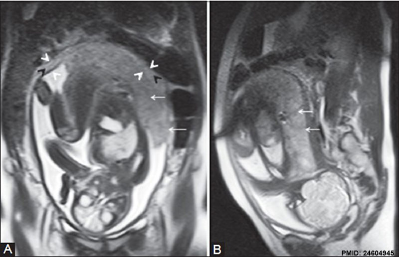 | 800 × 516 (60 KB) | Z8600021 (talk | contribs) | ==Placenta MRI (37 weeks)== Normal mature placenta at upper segment of uterus at 37 weeks of gestation. (A, B) Coronal and sagittal T2 HASTE MR images show a mildly heterogeneous placenta with normal placental septi (white arrows) and triple-layered ... |
You cannot overwrite this file.
File usage
The following page uses this file: