File:Pineal histology 001.jpg: Difference between revisions
From Embryology
No edit summary |
|||
| Line 1: | Line 1: | ||
==Pineal Histology== | ==Pineal Histology== | ||
Pineal gland, sheep H&E, pinealocytes | |||
Pin42he.jpg | Pin42he.jpg | ||
Revision as of 07:48, 30 September 2010
Pineal Histology
Pineal gland, sheep H&E, pinealocytes
Pin42he.jpg
Image Source: UWA Blue Histology http://www.lab.anhb.uwa.edu.au/mb140/CorePages/Endocrines/endocrin.htm
Links: Histology | Histology Stains | Blue Histology images copyright Lutz Slomianka 1998-2009. The literary and artistic works on the original Blue Histology website may be reproduced, adapted, published and distributed for non-commercial purposes. See also the page Histology Stains.
Cite this page: Hill, M.A. (2024, May 2) Embryology Pineal histology 001.jpg. Retrieved from https://embryology.med.unsw.edu.au/embryology/index.php/File:Pineal_histology_001.jpg
- © Dr Mark Hill 2024, UNSW Embryology ISBN: 978 0 7334 2609 4 - UNSW CRICOS Provider Code No. 00098G
File history
Click on a date/time to view the file as it appeared at that time.
| Date/Time | Thumbnail | Dimensions | User | Comment | |
|---|---|---|---|---|---|
| current | 16:00, 12 May 2012 | 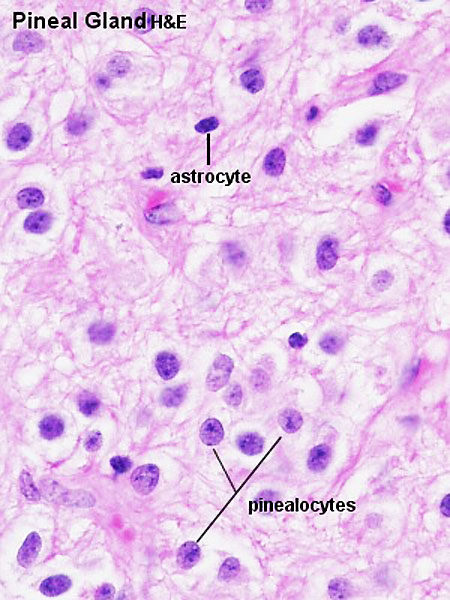 | 450 × 600 (75 KB) | Z8600021 (talk | contribs) | |
| 13:48, 4 October 2009 | 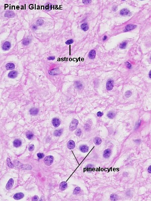 | 300 × 400 (47 KB) | S8600021 (talk | contribs) | Pin42he.jpg |
You cannot overwrite this file.