File:Ovary histology 061.jpg: Difference between revisions
(Z8600021 uploaded a new version of "File:Ovary histology 061.jpg") |
|||
| Line 6: | Line 6: | ||
** '''cumulus oophrous''' (Latin, ''cumulus'' = a little mound; Greek, ''oo''= egg, ''phorus''=carrying) granulosa cells directly around the zone pellucida and released with the oocyte. | ** '''cumulus oophrous''' (Latin, ''cumulus'' = a little mound; Greek, ''oo''= egg, ''phorus''=carrying) granulosa cells directly around the zone pellucida and released with the oocyte. | ||
** '''membrana granulosa''' (mural granulosa cell) granulosa cells forming the layer within the follicle antral wall. | ** '''membrana granulosa''' (mural granulosa cell) granulosa cells forming the layer within the follicle antral wall. | ||
** '''discus proligerus''' can refer to the attachment between cumulus oophrous and | ** '''discus proligerus''' can refer to the attachment between cumulus oophrous and membrana granulosa. | ||
* These spaces enlarge and fuse to form the '''follicular antrum''', which is the defining feature of the secondary follicle. | * These spaces enlarge and fuse to form the '''follicular antrum''', which is the defining feature of the secondary follicle. | ||
** antrum follicular fluid is released at along with the oocyte at ovulation. | ** antrum follicular fluid is released at along with the oocyte at ovulation. | ||
| Line 12: | Line 12: | ||
** note the many mitotic profiles visible in these cells. | ** note the many mitotic profiles visible in these cells. | ||
* The theca folliculi differentiates with the continued growth of the follicle into a '''theca interna''' and a '''theca externa'''. | * The theca folliculi differentiates with the continued growth of the follicle into a '''theca interna''' and a '''theca externa'''. | ||
** theca interna vascularization improves and the spindle-shaped or polyhedral cells in this layer start to produce estrogen (oestrogen). | ** theca interna vascularization improves and the spindle-shaped or polyhedral cells in this layer start to produce {{estrogen}} (oestrogen). | ||
** theca externa retains the characteristics of a highly cellular connective tissue with smooth muscle cells. | ** theca externa retains the characteristics of a highly cellular connective tissue with smooth muscle cells. | ||
* The oocyte of the secondary follicle reaches a diameter of about 125 µm. | * The oocyte of the secondary follicle reaches a diameter of about 125 µm. | ||
Latest revision as of 08:39, 19 March 2020
Ovary - Secondary Follicle
Histological image of a secondary (antral) stage of follicle development in the ovary.
- Small fluid-filled spaces become visible between the granulosa cells as the follicle reaches a diameter of about 400 µm.
- granulosa cells can also have specific names depending upon location within the follicle.
- cumulus oophrous (Latin, cumulus = a little mound; Greek, oo= egg, phorus=carrying) granulosa cells directly around the zone pellucida and released with the oocyte.
- membrana granulosa (mural granulosa cell) granulosa cells forming the layer within the follicle antral wall.
- discus proligerus can refer to the attachment between cumulus oophrous and membrana granulosa.
- These spaces enlarge and fuse to form the follicular antrum, which is the defining feature of the secondary follicle.
- antrum follicular fluid is released at along with the oocyte at ovulation.
- The oocyte is now located eccentrically in the follicle in the cumulus oophorus, where it is surrounded by granulosa cells.
- note the many mitotic profiles visible in these cells.
- The theca folliculi differentiates with the continued growth of the follicle into a theca interna and a theca externa.
- theca interna vascularization improves and the spindle-shaped or polyhedral cells in this layer start to produce estrogen (oestrogen).
- theca externa retains the characteristics of a highly cellular connective tissue with smooth muscle cells.
- The oocyte of the secondary follicle reaches a diameter of about 125 µm.
- The follicle itself reaches a diameter of about 10-15 mm.
There are several different nomenclatures for the stages of follicle maturation. It probably does not matter which naming system you use, as long as you are consistent and use the same set of terminology for all stages. Early stages of follicle development appear to be gonadotropin (Gn) independent and with development become gonadotropin "sensitive" and then "dependent" . (UK spelling is gonadotrophin).
- Primordial Follicle - Alternate nomenclature: small follicle or type 1, 2, 3 (25cells)
- Primary Follicle - Alternate nomenclature: preantral follicle or type 4 (26-100 cells), type 5 (101-300 cells)
- Secondary Follicle - Alternate nomenclature: small and large antral follicle or type 6 (3001-500 cells), type 7 (501-1000 cells)
- Preovulatory Follicle - Alternate nomenclature: Graafian follicle or type 8 (>1000 cells)
- Atresia - At any one time the majority of follicles are destined not to complete maturation and at any stage (from type 4-7) degeneration of the follicle can occur.
- Links: Oocyte Development | Zona pellucida | Granulosa cell | Ovary Development
Ovary histology: Tunica Albuginea x20 | Tunica albuginea, Germinal epithelium x40 | Primary follicle, primordial follicle, oocyte, x40 | Secondary follicle, cumulus oophorus, zona pelucida, granulosa cells, oocyte x20 | Corpus luteum, theca lutein cells, granulosa lutein cells, Loupe | Corpus luteum, theca lutein cells, granulosa lutein cells, x10 | Corpus luteum, theca lutein cells, granulosa lutein cells, x40 | Corpus albicans, primary follicle, primordial follicle, granulosa cells, oocyte x20 | Menstrual Cycle | Ovary Development
- Image Links: large 1000px | medium 800px | small 400px
File:Ovary_histology_006.jpg
Ovary, monkey H&E reproductive system, female, secondary follicle, cumulus oophorus, zona_pelucida,_granulosa_cells,_oocyte x20
Links: Histology | Histology Stains | Blue Histology images copyright Lutz Slomianka 1998-2009. The literary and artistic works on the original Blue Histology website may be reproduced, adapted, published and distributed for non-commercial purposes. See also the page Histology Stains.
Cite this page: Hill, M.A. (2024, April 26) Embryology Ovary histology 061.jpg. Retrieved from https://embryology.med.unsw.edu.au/embryology/index.php/File:Ovary_histology_061.jpg
- © Dr Mark Hill 2024, UNSW Embryology ISBN: 978 0 7334 2609 4 - UNSW CRICOS Provider Code No. 00098G
Cite this page: Hill, M.A. (2024, April 26) Embryology Ovary histology 061.jpg. Retrieved from https://embryology.med.unsw.edu.au/embryology/index.php/File:Ovary_histology_061.jpg
- © Dr Mark Hill 2024, UNSW Embryology ISBN: 978 0 7334 2609 4 - UNSW CRICOS Provider Code No. 00098G
File history
Click on a date/time to view the file as it appeared at that time.
| Date/Time | Thumbnail | Dimensions | User | Comment | |
|---|---|---|---|---|---|
| current | 17:52, 27 December 2014 | 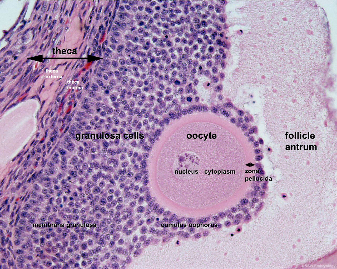 | 1,280 × 1,024 (438 KB) | Z8600021 (talk | contribs) | addd theca layer label |
| 21:37, 5 May 2011 | 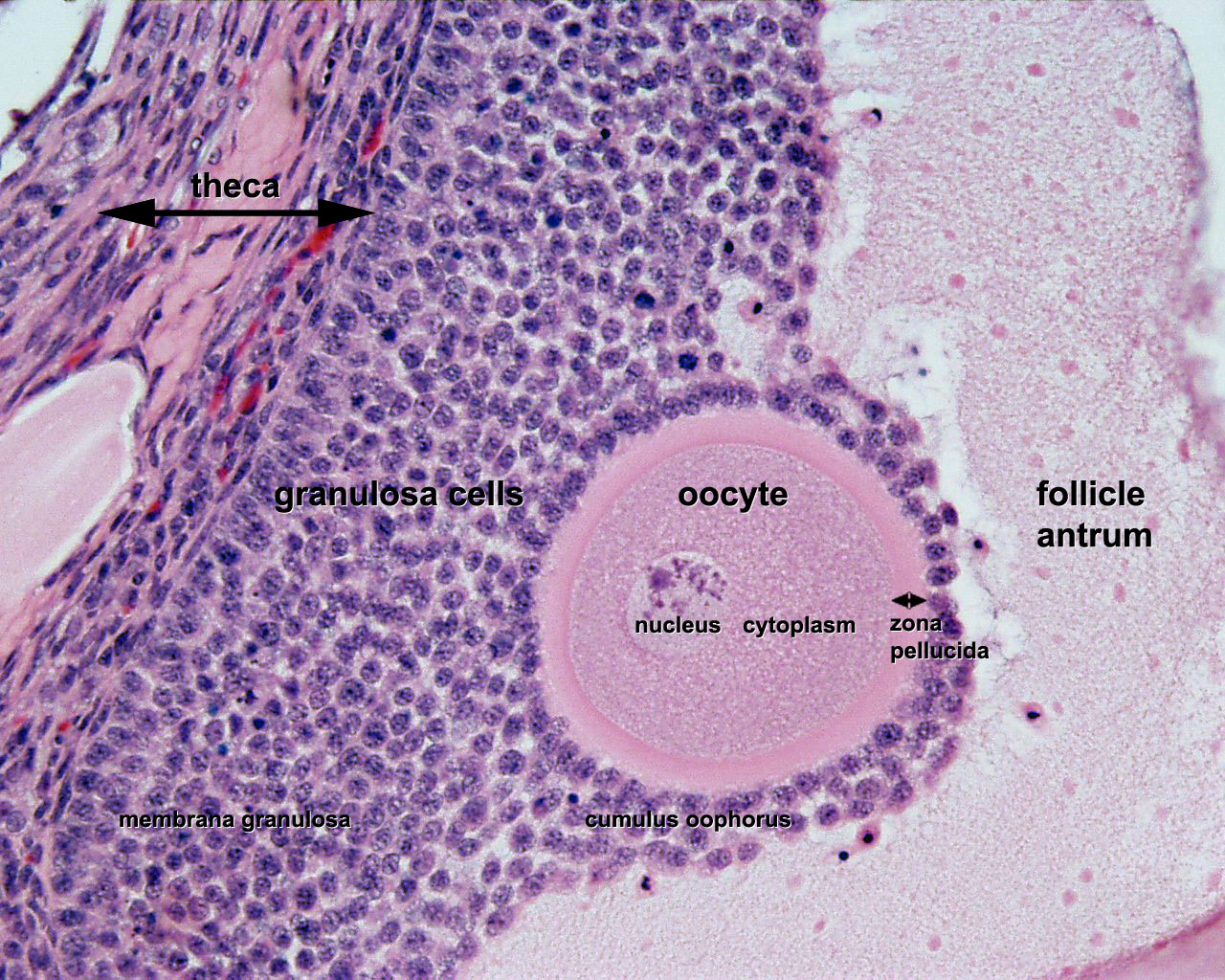 | 1,280 × 1,024 (436 KB) | S8600021 (talk | contribs) | ==Ovary - Secondary Follicle== * Small fluid-filled spaces become visible between the granulosa cells as the follicle reaches a diameter of about 400 µm. * These spaces enlarge and fuse to form the follicular antrum, which is the defining feature of th |
You cannot overwrite this file.
File usage
The following 9 pages use this file: