File:Ovary histology 005.jpg
Original file (1,280 × 1,024 pixels, file size: 354 KB, MIME type: image/jpeg)
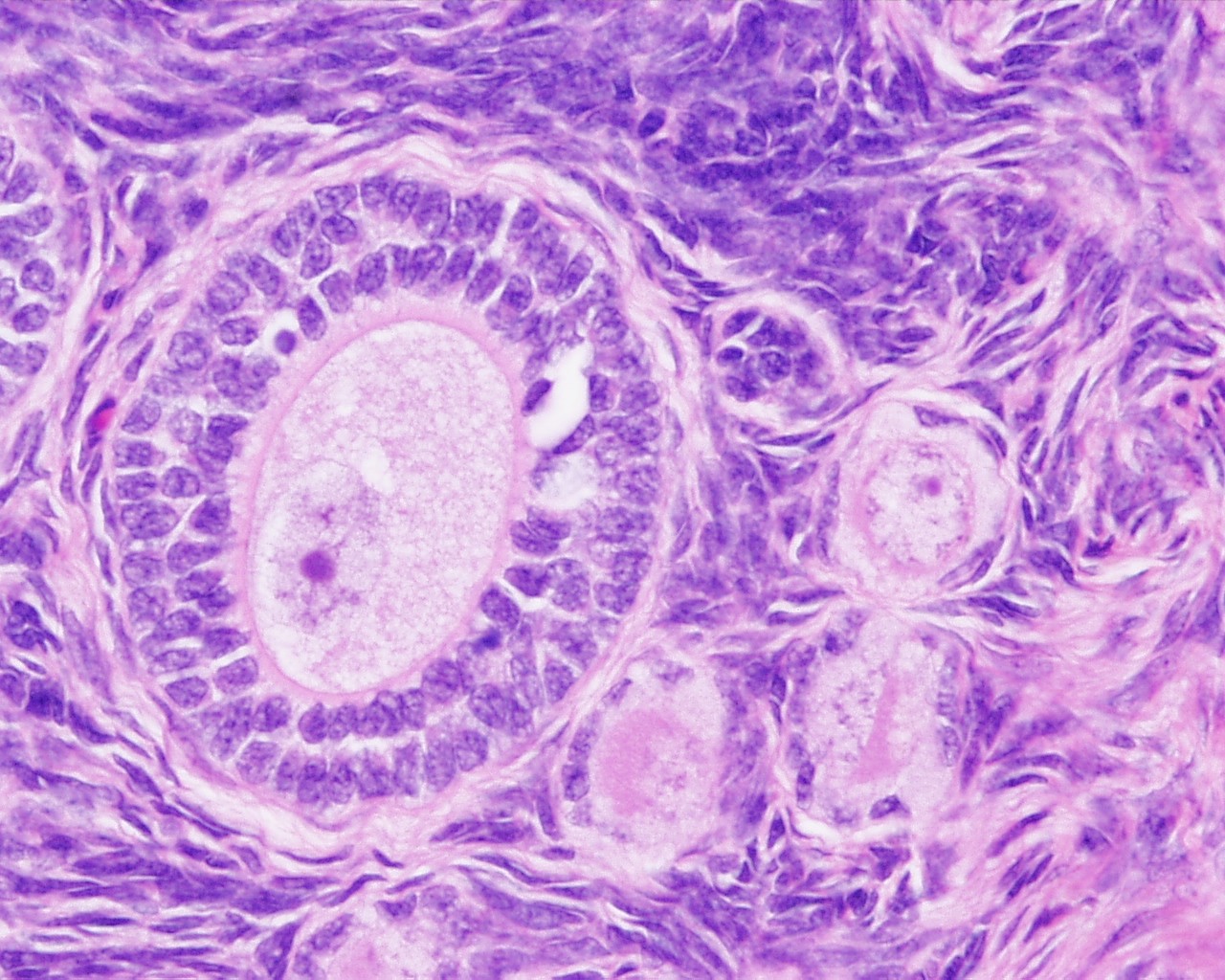
Ovary - Primary Follicle
Primordial follicles are located in the cortex just beneath tunica albuginea. One layer of flattened follicular cells surround the oocyte (about 30 µm in diameter). The nucleus of the oocyte is positioned eccentric in the cell. It appears very light and contains a prominent nucleolus. Most organelles of the oocyte aggregate in the centre of the cell, where they form the vitelline body (probably not visible in any of the available preparations).
Primary follicle is the first morphological stage that marks the onset of follicular maturation (Which hormone stimulates follicular maturation and where is this hormone produced?). The previously flattened cell surrounding the oocyte now form a cuboidal or columnar epithelium surrounding the oocyte. Their cytoplasm may have a granular appearance, and they are for this reason also called granulosa cells. The continued proliferation of these cells will result in the formation of a stratified epithelium (with a distinct basement membrane) surrounding the oocyte. The zona pellucida (glycoproteins between interdigitating processes of oocyte and granulosa cells) becomes visible. Parenchymal cells of the ovary surrounding the growing follicle become organised in concentric sheaths, the theca folliculi.
File:Ovary_histology_005.jpg
Ovary, monkey H&E reproductive system, female, primary follicle, primordial follicle, oocyte, x40
Ovary histology: Tunica Albuginea x20 | Tunica albuginea, Germinal epithelium x40 | Primary follicle, primordial follicle, oocyte, x40 | Secondary follicle, cumulus oophorus, zona pelucida, granulosa cells, oocyte x20 | Corpus luteum, theca lutein cells, granulosa lutein cells, Loupe | Corpus luteum, theca lutein cells, granulosa lutein cells, x10 | Corpus luteum, theca lutein cells, granulosa lutein cells, x40 | Corpus albicans, primary follicle, primordial follicle, granulosa cells, oocyte x20 | Menstrual Cycle | Ovary Development
Links: Histology | Histology Stains | Blue Histology images copyright Lutz Slomianka 1998-2009. The literary and artistic works on the original Blue Histology website may be reproduced, adapted, published and distributed for non-commercial purposes. See also the page Histology Stains.
Cite this page: Hill, M.A. (2024, April 27) Embryology Ovary histology 005.jpg. Retrieved from https://embryology.med.unsw.edu.au/embryology/index.php/File:Ovary_histology_005.jpg
- © Dr Mark Hill 2024, UNSW Embryology ISBN: 978 0 7334 2609 4 - UNSW CRICOS Provider Code No. 00098G
File history
Click on a date/time to view the file as it appeared at that time.
| Date/Time | Thumbnail | Dimensions | User | Comment | |
|---|---|---|---|---|---|
| current | 21:10, 23 February 2011 | | 1,280 × 1,024 (354 KB) | S8600021 (talk | contribs) | File:Ovary_histology_005.jpg Ovary,_monkey_H&E_reproductive_system,_female,_primary_follicle,_primordial_follicle,_oocytex40.jpg {{Ovary Histology}} {{Template:Blue Histology}} Category:Monkey Category:Genital Category:Histology [[Catego |
You cannot overwrite this file.
File usage
The following 5 pages use this file: