File:Ovary- atretic follicle 01.jpg: Difference between revisions
From Embryology
No edit summary |
mNo edit summary |
||
| Line 1: | Line 1: | ||
Ovary | ==Ovary Atretic Follicle== | ||
Cat ovary {{HE}} | |||
UNSW Anatomy slide collection | * Note the follicular fluid space is "collapsed" and not a smooth "inflated" space. Follicle wall, granulosa cell layer, is also folded. | ||
* White space within follicular fluid space is histological artefact, shrinkage. | |||
:'''Links:''' [[:File:Ovary- atretic follicle 02.jpg|small image]] | [[:File:Ovary- atretic follicle 01.jpg|Large image]] | [[Ovary Development]] | |||
---- | |||
low magnification x10, UNSW Anatomy slide collection | |||
[[Category:Ovary]] [[Category:Histology]] [[Category:Cat]] | [[Category:Ovary]] [[Category:Histology]] [[Category:Cat]] | ||
Latest revision as of 11:48, 9 May 2013
Ovary Atretic Follicle
Cat ovary (Stain - Haematoxylin Eosin)
- Note the follicular fluid space is "collapsed" and not a smooth "inflated" space. Follicle wall, granulosa cell layer, is also folded.
- White space within follicular fluid space is histological artefact, shrinkage.
- Links: small image | Large image | Ovary Development
low magnification x10, UNSW Anatomy slide collection
File history
Click on a date/time to view the file as it appeared at that time.
| Date/Time | Thumbnail | Dimensions | User | Comment | |
|---|---|---|---|---|---|
| current | 08:17, 10 May 2010 | 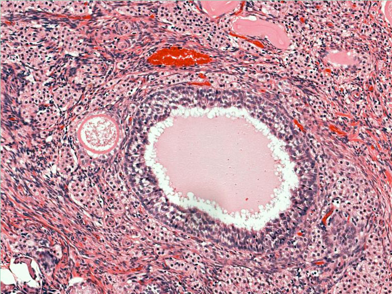 | 793 × 595 (225 KB) | S8600021 (talk | contribs) | Ovary - atretic follicle (cat) x10 magnification UNSW Anatomy slide collection |
You cannot overwrite this file.
File usage
The following page uses this file: