File:Oesophagus histology 03.jpg: Difference between revisions
From Embryology
No edit summary |
No edit summary |
||
| Line 3: | Line 3: | ||
* note the difference in this epithelium in comparison to the skin epithelium. | * note the difference in this epithelium in comparison to the skin epithelium. | ||
* the surface cell layers still have their nuclei. | * the surface cell layers still have their nuclei. | ||
{{Oesophagus Histology}} | |||
Latest revision as of 12:54, 26 March 2012
Oesophagus Histology
- note the difference in this epithelium in comparison to the skin epithelium.
- the surface cell layers still have their nuclei.
- Oesophagus Histology: Skeletal and Smooth Muscle | Submucosa Gland | Muscle | Gland-Muscle Animation | Epithelia and Lamina Propria | Labeled Epithelia | Labeled Connective Tissue | Unlabelled | Unlabelled | Oesophagus Development
Links: Histology | Histology Stains | Blue Histology images copyright Lutz Slomianka 1998-2009. The literary and artistic works on the original Blue Histology website may be reproduced, adapted, published and distributed for non-commercial purposes. See also the page Histology Stains.
Cite this page: Hill, M.A. (2024, May 15) Embryology Oesophagus histology 03.jpg. Retrieved from https://embryology.med.unsw.edu.au/embryology/index.php/File:Oesophagus_histology_03.jpg
- © Dr Mark Hill 2024, UNSW Embryology ISBN: 978 0 7334 2609 4 - UNSW CRICOS Provider Code No. 00098G
File history
Click on a date/time to view the file as it appeared at that time.
| Date/Time | Thumbnail | Dimensions | User | Comment | |
|---|---|---|---|---|---|
| current | 17:39, 5 March 2012 | 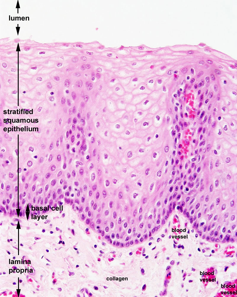 | 800 × 1,000 (209 KB) | Z8600021 (talk | contribs) | ==Oesophagus Histology== {{Blue Histology}} Category:Histology Category:Gastrointestinal Tract |
You cannot overwrite this file.
File usage
The following 7 pages use this file: