File:Oesophagus MALT.jpg: Difference between revisions
From Embryology
No edit summary |
|||
| Line 1: | Line 1: | ||
==Oesophagus Mucosa Associated Lymphoid Tissue== | ==Oesophagus - Mucosa Associated Lymphoid Tissue== | ||
* MALT - Mucosa Associated Lymphoid Tissue | * MALT - Mucosa Associated Lymphoid Tissue | ||
{{Immune Images 2}} | |||
{{Blue Histology}} | {{Blue Histology}} | ||
Revision as of 10:46, 24 February 2012
Oesophagus - Mucosa Associated Lymphoid Tissue
- MALT - Mucosa Associated Lymphoid Tissue
- Immune Images: Oesophagus MALT | Colon MALT | Peyer's patch overview | Peyer's patch detail | Cartoon - IEL development | Cartoon - IEL function | Cartoon - IEL differentiation | Mesenteric Lymph Nodes overview | Palatine Tonsil | Tonsil | Immune System Development
Links: Histology | Histology Stains | Blue Histology images copyright Lutz Slomianka 1998-2009. The literary and artistic works on the original Blue Histology website may be reproduced, adapted, published and distributed for non-commercial purposes. See also the page Histology Stains.
Cite this page: Hill, M.A. (2024, May 7) Embryology Oesophagus MALT.jpg. Retrieved from https://embryology.med.unsw.edu.au/embryology/index.php/File:Oesophagus_MALT.jpg
- © Dr Mark Hill 2024, UNSW Embryology ISBN: 978 0 7334 2609 4 - UNSW CRICOS Provider Code No. 00098G
Original file name: Oes04he.jpg
File history
Click on a date/time to view the file as it appeared at that time.
| Date/Time | Thumbnail | Dimensions | User | Comment | |
|---|---|---|---|---|---|
| current | 10:09, 24 February 2012 | 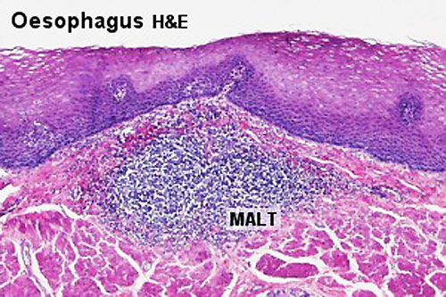 | 500 × 333 (73 KB) | Z8600021 (talk | contribs) | |
| 08:17, 24 December 2010 |  | 300 × 200 (36 KB) | S8600021 (talk | contribs) | ==Mucosa Associated Lymphoid Tissue== (MALT) Original file name: Oes04he.jpg Category:Immune Category:Histology Category:Gastrointestinal tract |
You cannot overwrite this file.