File:ORahilly1987 fig20-2.jpg: Difference between revisions
ORahilly1987_fig20-2.jpg (600 × 549 pixels, file size: 46 KB, MIME type: image/jpeg)
mNo edit summary |
mNo edit summary |
||
| (4 intermediate revisions by the same user not shown) | |||
| Line 1: | Line 1: | ||
==Fig. 20-2. Head Superficial Vascular Plexus - Stages 20-23== | ==Fig. 20-2. Head Superficial Vascular Plexus - Stages 20-23== | ||
Composite drawing showing the edge of the superficial vascular plexus in the head at stages 20-23. A subcutaneous capillary plexus that spreads upward toward the vertex of the head is a conspicuous feature of specimens measuring from about 20-24 mm in length. | Composite drawing showing the edge of the superficial vascular plexus in the head at stages 20-23. | ||
A subcutaneous capillary plexus that spreads upward toward the vertex of the head is a conspicuous feature of specimens measuring from about 20-24 mm in length. | |||
The transition from vascular to avascular mesoderm is more gradual in earlier stages and becomes more abrupt later. | The transition from vascular to avascular mesoderm is more gradual in earlier stages and becomes more abrupt later. | ||
| Line 7: | Line 9: | ||
Numbers refer to specific Carnegie embryos. | Numbers refer to specific Carnegie embryos. | ||
:'''Links:''' [[Week 8]] | [[Carnegie stage 20]] | [[Carnegie stage 21]] | [[Carnegie stage 22]] | [[Carnegie stage 23]] | |||
<br> | |||
{{Carnegie stage 20 links}} | |||
{{Carnegie stage 21 links}} | |||
{{Carnegie stage 22 links}} | |||
{{Carnegie stage 23 links}} | |||
<br> | |||
{{ORahilly1987 figures}} | {{ORahilly1987 figures}} | ||
[[Category:Head]][[Category:Human Embryo]][[Category:Week 8]] | [[Category:Head]][[Category:Human Embryo]][[Category:Cardiovascular]][[Category:Week 8]] | ||
[[Category:Carnegie Stage 20]][[Category:Carnegie Stage 21]][[Category:Carnegie Stage 22]][[Category:Carnegie Stage 23]] | [[Category:Carnegie Stage 20]][[Category:Carnegie Stage 21]][[Category:Carnegie Stage 22]][[Category:Carnegie Stage 23]] | ||
[[Category:Carnegie Embryo 2937]][[Category:Carnegie Embryo 966]] | |||
Latest revision as of 13:28, 22 May 2017
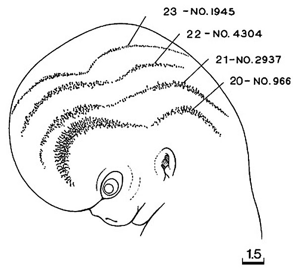
Fig. 20-2. Head Superficial Vascular Plexus - Stages 20-23
Composite drawing showing the edge of the superficial vascular plexus in the head at stages 20-23.
A subcutaneous capillary plexus that spreads upward toward the vertex of the head is a conspicuous feature of specimens measuring from about 20-24 mm in length.
The transition from vascular to avascular mesoderm is more gradual in earlier stages and becomes more abrupt later.
Numbers refer to specific Carnegie embryos.
- Links: Week 8 | Carnegie stage 20 | Carnegie stage 21 | Carnegie stage 22 | Carnegie stage 23
| Stage 22 Links: Week 8 | System Development | Lecture - Limb | Lecture - Head Development | Lecture - Sensory | Science Practical - Head | Science Practical - Sensory | Science Practical - Urogenital | Historic - Skull Development | Carnegie Embryos | Madrid Embryos | Category:Carnegie Stage 22 | Next Stage 23 |
| Week 8, GA week 10, 54 - 56 days, CRL 23 - 28 mm, Carnegie Embryos |
| Historic Papers: 1914 | 1954 Stage 19-23 |
- 1987 Stages: Introduction | 1 | 2 | 3 | 4 | 5 | 6 | 7 | 8 | 9 | 10 | 11 | 12 | 13 | 14 | 15 | 16 | 17 | 18 | 19 | 20 | 21 | 22 | 23 | References | Appendix 1 | Appendix 2 | Historic Papers | Embryonic Development
Cite this page: Hill, M.A. (2024, April 26) Embryology ORahilly1987 fig20-2.jpg. Retrieved from https://embryology.med.unsw.edu.au/embryology/index.php/File:ORahilly1987_fig20-2.jpg
- © Dr Mark Hill 2024, UNSW Embryology ISBN: 978 0 7334 2609 4 - UNSW CRICOS Provider Code No. 00098G
File history
Click on a date/time to view the file as it appeared at that time.
| Date/Time | Thumbnail | Dimensions | User | Comment | |
|---|---|---|---|---|---|
| current | 18:52, 17 July 2015 | | 600 × 549 (46 KB) | Z8600021 (talk | contribs) |
You cannot overwrite this file.
File usage
The following page uses this file: